. 2015 Aug 19;589(18):2297-303.
doi: 10.1016/j.febslet.2015.07.009. Epub 2015 Jul 18.

Recognition of xyloglucan by the crystalline cellulose-binding site of a family 3a carbohydrate-binding module

Affiliations

Recognition of xyloglucan by the crystalline cellulose-binding site of a family 3a carbohydrate-binding module

Mercedes C Hernandez-Gomez et al. FEBS Lett. .

Abstract

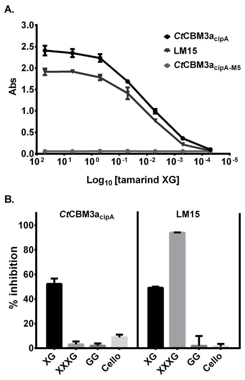
Type A non-catalytic carbohydrate-binding modules (CBMs), exemplified by CtCBM3acipA, are widely believed to specifically target crystalline cellulose through entropic forces. Here we have tested the hypothesis that type A CBMs can also bind to xyloglucan (XG), a soluble β-1,4-glucan containing α-1,6-xylose side chains. CtCBM3acipA bound to xyloglucan in cell walls and arrayed on solid surfaces. Xyloglucan and cellulose were shown to bind to the same planar surface on CBM3acipA. A range of type A CBMs from different families were shown to bind to xyloglucan in solution with ligand binding driven by enthalpic changes. The nature of CBM-polysaccharide interactions is discussed.

Keywords: CBM3a; Carbohydrate-binding module; Crystalline cellulose; Plant cell wall; Xyloglucan.


Figures

Fig. 1
Fig. 1
Indirect immunofluorescence detection of crystalline cellulose and xyloglucan in sections of xyloglucan-rich tamarind seed cotyledon parenchyma and cellulose-rich cotton fibres, respectively. Calcofluor White staining indicates location of all cell walls. LM15 binding to cell walls of tamarind cotyledon parenchyma cells and primary cell walls of cotton fibres (arrows) is abolished after a xyloglucanase pre-treatment. CtCBM3acipA WT binding to cell walls of tamarind cotyledon parenchyma cells is abolished by xyloglucanase pre-treatment however its binding to the secondary cell wall of cotton fibres is not. CtCBM3acipA-M5 did not bind to cell walls of tamarind cotyledon parenchyma cells nor to cotton fibre secondary cell walls.
Fig. 2
Fig. 2
In vitro recognition of xyloglucan. A. ELISA analysis of CtCBM3acipA, CtCBM3acipA-M5 and LM15 binding to tamarind xyloglucan. B. Competitive-inhibition ELISAs of LM15 and CtCBM3acipA binding to immobilised xyloglucan with xyloglucan (XG), xyloglucan heptasaccharide (XXXG), guar galactomannan (GG) and cellohexaose (Cello) in the soluble phase at 25 μg/ml. Y axis represents the percentage of inhibition achieved by the polysaccharides/haptens. Error bars: SD (n=3)
Fig. 3
Fig. 3
Affinity gel electrophoresis of type A CBMs. The proteins were electrophoresed in gels containing 0.1% xyloglucan, 0.1% barley β1,3-β1,4-mixed linkage glucan (β-glucan) or no glucan (control). ~10 μg of protein was loaded in each well. CBM65, which binds to a range of β-glucans was used as a positive control and GFP and BSA were deployed as non-binding negative controls.
Fig. 4
Fig. 4
Representative isothermal titration calorimetry (ITC) data of CtCBM3acipA-M5 titrated with carbohydrates. The soluble ligand (10 mg/mL in the syringe) was titrated into 54 μM CtCBM3acipA-M5 in the cell. In the case of cellulose the CBM was titrated into cellulose in the cell. The top half of each titration shows the raw injection heats; the bottom half, the integrated peak areas fitted using a single-site model (MicroCal Origin v7.0). ITC was carried out in 50 mM Na-HEPES pH 7.5 at 25°C.

References

    1. Gilbert HJ. The biochemistry and structural biology of plant cell wall deconstruction. Plant Physiol. 2010;153:444–455. - PMC - PubMed
    1. Lombard V, Golaconda Ramulu H, Drula E, Coutinho PM, Henrissat B. The carbohydrate-active enzymes database (CAZy) in 2013. Nucleic acids research. 2014;42:D490–495. - PMC - PubMed
    1. Gilbert HJ, Knox JP, Boraston AB. Advances in understanding the molecular basis of plant cell wall polysaccharide recognition by carbohydrate-binding modules. Curr Opin Struct Biol. 2013;23:669–677. - PubMed
    1. Boraston AB, Bolam DN, Gilbert HJ, Davies GJ. Carbohydrate-binding modules: fine-tuning polysaccharide recognition. Biochem J. 2004;382:769–781. - PMC - PubMed
    1. Raghothama S, Simpson PJ, Szabo L, Nagy T, Gilbert HJ, Williamson MP. Solution structure of the CBM10 cellulose binding module from Pseudomonas xylanase A. Biochemistry. 2000;39:978–984. - PubMed

Publication types