

Magnetic resonance imaging in Alzheimer's Disease Neuroimaging Initiative 2
- PMID: 26194310
- PMCID: PMC4523217
- DOI: 10.1016/j.jalz.2015.05.002
Magnetic resonance imaging in Alzheimer's Disease Neuroimaging Initiative 2
Abstract
Introduction: Alzheimer's Disease Neuroimaging Initiative (ADNI) is now in its 10th year. The primary objective of the magnetic resonance imaging (MRI) core of ADNI has been to improve methods for clinical trials in Alzheimer's disease (AD) and related disorders.
Methods: We review the contributions of the MRI core from present and past cycles of ADNI (ADNI-1, -Grand Opportunity and -2). We also review plans for the future-ADNI-3.
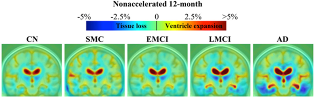
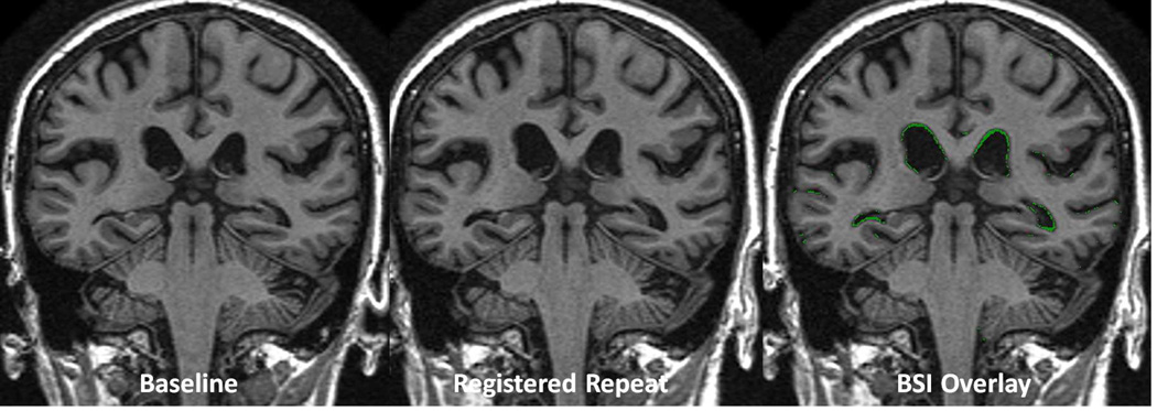
Results: Contributions of the MRI core include creating standardized acquisition protocols and quality control methods; examining the effect of technical features of image acquisition and analysis on outcome metrics; deriving sample size estimates for future trials based on those outcomes; and piloting the potential utility of MR perfusion, diffusion, and functional connectivity measures in multicenter clinical trials.
Discussion: Over the past decade the MRI core of ADNI has fulfilled its mandate of improving methods for clinical trials in AD and will continue to do so in the future.
Keywords: ADNI; Alzheimer's Disease Neuroimaging Initiative; Alzheimer's disease; Diffusion; MRI; Neuroimaging; Perfusion; Resting functional MRI.
Copyright © 2015 The Authors. Published by Elsevier Inc. All rights reserved.
Figures
References
-
- Rydberg JN, Riederer SJ, Rydberg CH, Jack CR. Contrast optimization of fluid-attenuated inversion recovery (FLAIR) imaging. Magn Reson Med. 1995;34(6):868–877. - PubMed
-
- Mueller S, Yushkevich P, Wang L, Van Leemput K, Mezher A, Iglesias JE, Das S, Weiner M. Collaboration for a systematic comparison of different techniques to measure subfield volumes: Announcement and first results. Alzheimer's & Dementia: The Journal of the Alzheimer's Association. 2013;9(4):P51.
Publication types
MeSH terms
Substances
Grants and funding
LinkOut - more resources
Full Text Sources
Other Literature Sources
Medical
Miscellaneous
