. 2015 Aug;29(8):737-56.
doi: 10.1007/s10822-015-9858-z. Epub 2015 Jul 21.

Modeling ligand recognition at the P2Y12 receptor in light of X-ray structural information

Affiliations

Modeling ligand recognition at the P2Y12 receptor in light of X-ray structural information

Silvia Paoletta et al. J Comput Aided Mol Des. 2015 Aug.

Abstract

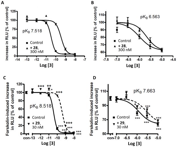
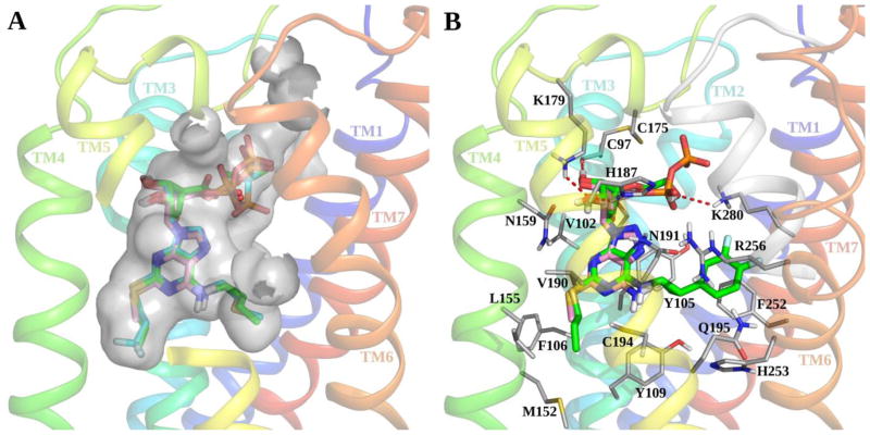
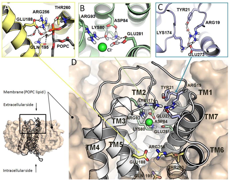
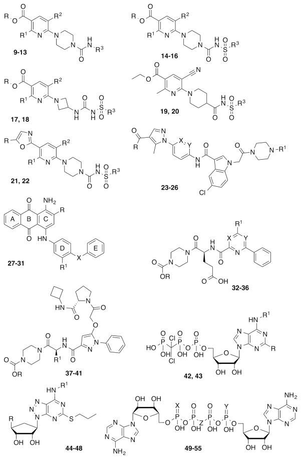
The G protein-coupled P2Y12 receptor (P2Y12R) is an important antithrombotic target and of great interest for pharmaceutical discovery. Its recently solved, highly divergent crystallographic structures in complex either with nucleotides (full or partial agonist) or with a nonnucleotide antagonist raise the question of which structure is more useful to understand ligand recognition. Therefore, we performed extensive molecular modeling studies based on these structures and mutagenesis, to predict the binding modes of major classes of P2Y12R ligands previously reported. Various nucleotide derivatives docked readily to the agonist-bound P2Y12R, but uncharged nucleotide-like antagonist ticagrelor required a hybrid receptor resembling the agonist-bound P2Y12R except for the top portion of TM6. Supervised molecular dynamics (SuMD) of ticagrelor binding indicated interactions with the extracellular regions of P2Y12R, defining possible meta-binding sites. Ureas, sulfonylureas, sulfonamides, anthraquinones and glutamic acid piperazines docked readily to the antagonist-bound P2Y12R. Docking dinucleotides at both agonist- and antagonist-bound structures suggested interactions with two P2Y12R pockets. Thus, our structure-based approach consistently rationalized the main structure-activity relationships within each ligand class, giving useful information for designing improved ligands.


Figures

Figure 1
Figure 1
Comparison between agonist- and antagonist-bound P2Y12R crystal structures. Side view of the crystallographic poses of (A) compound 3 (pink carbon sticks) at the agonist-bound P2Y12R structure and (B) compound 19 (green carbon sticks) at the antagonist-bound P2Y12R structure. Side chains of residues important for ligand recognition are shown in thin sticks (grey carbons) and polar interactions are indicated by red dashed lines. Non-polar hydrogen atoms are not displayed. 2D representation of the binding modes of (C) 3 and (D) 19 at the P2Y12R. Magenta arrows indicate H-bonds, red-purple lines indicate salt bridges and green lines indicate π-π stacking interactions. Residues are colored based on their features: positively charged residues in purple, polar residues in cyan and hydrophobic residues in green. (E) RMSD in Å of the residues located within 3 Å from the crystallographic poses of compounds 3 and 19 between the two P2Y12R crystal structures. Residues whose RMSD values are not reported are not resolved in the antagonist-bound P2Y12R structure.
Figure 2
Figure 2
Docking of P2Y12R agonists to the agonist-bound crystal structure: Side view of the docking poses of nucleoside 5′-diphosphates 1 (cyan carbon sticks) and 4 (magenta carbons sticks) and crystal pose of compound 3 (pink carbon sticks) at the agonist-bound P2Y12R structure. Non-polar hydrogen atoms are not displayed. The surface of the binding site is shown in grey. The view of TM5 is partially omitted.
Figure 3
Figure 3
Docking of urea, sulfonylurea, sulfonamide and amide derivatives to the antagonist-bound P2Y12R crystal structure. (A) Side view of the docking poses of compounds 13 (cyan carbon sticks), 18 (magenta carbons sticks) and 22 (pink carbons sticks) and crystal pose of compound 19 (green carbon sticks) at the antagonist-bound P2Y12R structure. (B) Side view of the docking pose of compound 26 (magenta carbons sticks) and crystal pose of compound 19 (green carbon sticks) at the antagonist-bound P2Y12R structure. Side chains of residues important for ligand recognition are shown in thin sticks (grey carbons), and polar interactions are indicated by red, dashed lines. Non-polar hydrogen atoms are not displayed.
Figure 4
Figure 4
Docking of anthraquinone and glutamic acid piperazine derivatives to the antagonist-bound P2Y12R crystal structure. (A) Side view of the docking pose of compound 27 (magenta carbon sticks) and crystal pose of compound 19 (green carbon sticks) at the antagonist-bound P2Y12R structure. (B) Side view of the docking poses of compounds 32 (magenta carbon sticks) and 41 (cyan carbon sticks) at the antagonist-bound P2Y12R crystal structure. Side chains of residues important for ligand recognition are shown in thin sticks (grey carbons), and red dashed lines indicate polar interactions formed by the docked compounds. Non-polar hydrogen atoms are not displayed.
Figure 5
Figure 5
Effects of anthraquinones (A), (B) 28 and (C), (D) 29 on compound 3-mediated inhibition of forskolin-induced cAMP response element (CRE)-driven luciferase expression in CHO Flp-In cells expressing the hP2Y12R fused to ECFP (panels A and C) or K280A-mutant receptor fused to ECFP (panels B and D). Means ± S.E. of 4 to 24 experiments.
Figure 6
Figure 6
Docking of nucleotide and nucleoside derivatives to the agonist-bound P2Y12R crystal structure and P2Y12R hybrid model. (A) Side view of the docking poses of compounds 43 (cyan carbon sticks) and 44 (green carbons sticks) and crystal pose of compound 3 (pink carbon sticks) at the agonist-bound P2Y12R structure. Non-polar hydrogen atoms are not displayed. The surface of the binding site is shown in grey. The view of TM5 is partially omitted. (B) Side view of the docking pose of compound 47 (green carbon sticks) at the P2Y12R hybrid model. Side chains of residues important for ligand recognition are shown in thin sticks (grey carbons) and polar interactions formed by compound 47 are indicated by red, dashed lines. Non-polar hydrogen atoms are not displayed. The crystal pose of compound 3 (pink carbon sticks) and the position of TM6 (white ribbon) in the agonist-bound P2Y12R structure are shown for comparison.
Figure 7
Figure 7
Docking of dinucleotides to the antagonist- and agonist-bound P2Y12R crystal structures. (A) Top view of the docking pose of compound 53 (cyan carbon sticks) at the antagonist-bound P2Y12R structure. (B) Top view of the docking pose of compound 49 (magenta carbon sticks) at the agonist-bound P2Y12R structure obtained after Induced Fit Docking. The crystal pose of compound 3 (pink carbon sticks) is shown for comparison. Side chains of residues important for ligand recognition are shown in thin sticks (grey carbons), and red dashed lines indicate polar interactions formed by the docked compounds. Non-polar hydrogen atoms are not displayed.
Figure 8
Figure 8
(A–C) Molecular details of key receptor-solvent/membrane interactions in P2Y12R prior to ligand recognition. Side chains of residues involved in key interaction networks are shown in sticks. Non-polar hydrogen atoms are not displayed. H-bonds and salt bridges are highlighted as dashed lines. Ions are depicted as spheres. (D) Overview of the apo-P2Y12R hybrid model in a membrane-like environment (POPC lipids).
Figure 9
Figure 9
(A), (D) Overview of identified ligand-receptor bound states that chronologically anticipate the orthosteric binding site recognition of compound 47 at P2Y12R. Ligand is depicted as a colored arrow. Arrow coloring scheme enables distinction of the single independent SuMD simulations that generated depicted bound states (SuMD seeds). Receptor ribbon representation is viewed from the membrane side facing TM5 and TM6. (B), (C) Atomistic level detailed representation of characterized convergent metastable-states during the approaching of compound 47 (yellow carbon sticks) to the P2Y12R from the extracellular binding pocket vestibule. Side chains of residues within 4 Å from compound 47 are highlighted (white carbon sticks). H-bonds are highlighted as yellow dashed lines. Hydrogen atoms are not displayed.
Figure 10
Figure 10
Comparison of the binding modes of different ligand classes at the P2Y12R. Superposition of the crystal poses of compounds 3 (pink carbon sticks) and 19 (green carbon sticks) and the docking poses of compounds 27 (magenta carbon sticks), 32 (blue carbon sticks), 43 (cyan carbon sticks) and 47 (dark green carbon sticks) at the P2Y12R. The antagonist-bound P2Y12R structure is shown in cyan ribbons. The view of TM5 is omitted. Similarities in the binding mode of different ligands are described.
Chart 1
Chart 1
Structures of representative nucleotides as P2Y12R agonists and partial agonists (compounds 18).
Chart 2
Chart 2
Structures of representative, reversible P2Y12R antagonists (nonnucleotides 941 and nucleotide-like 4255) belonging to different chemical classes. Substituents, references and available pharmacological data at the hP2Y12R are reported in Table 1.
Chart 3
Chart 3
Structures of two irreversible P2Y12R antagonist prodrugs (thienopyridines 56 and 57).

References

    1. Abbracchio MP, Burnstock G, Boeynaems JM, Barnard EA, Boyer JL, Kennedy C, Knight GE, Fumagalli M, Gachet C, Jacobson KA, Weisman GA. International Union of Pharmacology LVIII: update on the P2Y G protein-coupled nucleotide receptors: from molecular mechanisms and pathophysiology to therapy. Pharmacol Rev. 2006;58:281–341. - PMC - PubMed
    1. Burnstock G, Knight GE. Cellular distribution and functions of P2 receptor subtypes in different systems. Int Rev Cytol. 2004;240:31–304. - PubMed
    1. Hollopeter G, Jantzen HM, Vincent D, Li G, England L, Ramakrishnan V, Yang RB, Nurden P, Nurden A, Julius D, Conley PB. Identification of the platelet ADP receptor targeted by antithrombotic drugs. Nature. 2001;409:202–207. - PubMed
    1. Hourani SM, Cusack NJ. Pharmacological receptors on blood platelets. Pharmacol Rev. 1991;43:243–98. - PubMed
    1. Jacobson KA, Deflorian F, Mishra S, Costanzi S. Pharmacochemistry of the platelet purinergic receptors. Purinergic Signal. 2011;7:305–324. - PMC - PubMed

Publication types

MeSH terms

LinkOut - more resources