. 2015 Jul 21;112(29):8922-8.
doi: 10.1073/pnas.1505464112.

Stem cells are units of natural selection for tissue formation, for germline development, and in cancer development

Affiliations

Stem cells are units of natural selection for tissue formation, for germline development, and in cancer development

Irving L Weissman. Proc Natl Acad Sci U S A. .

Abstract

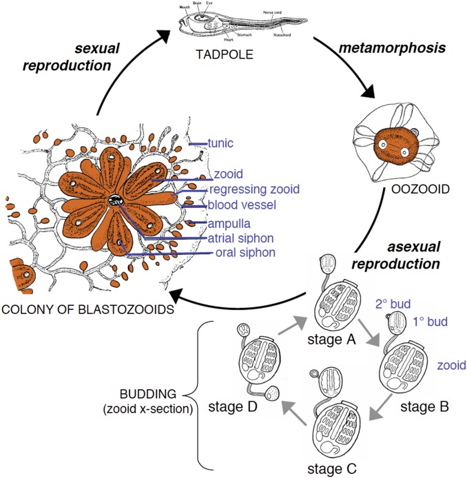
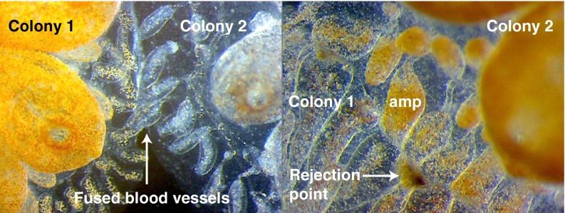
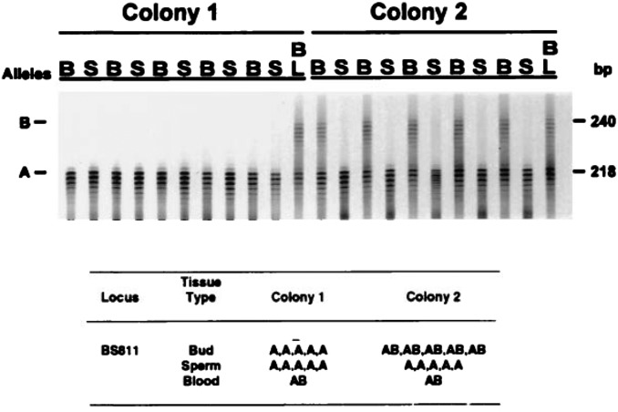
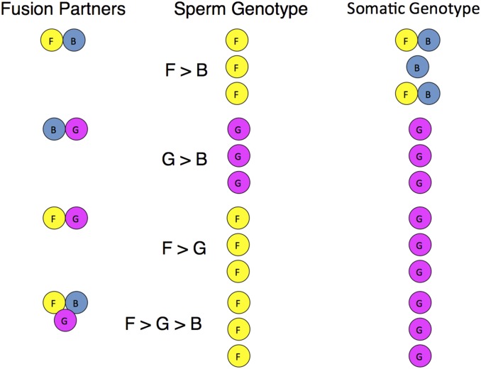
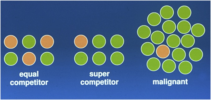
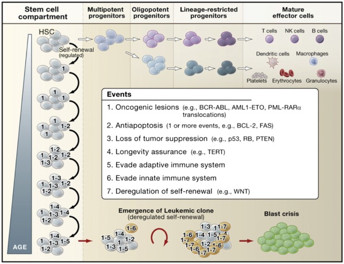
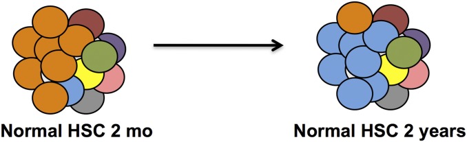
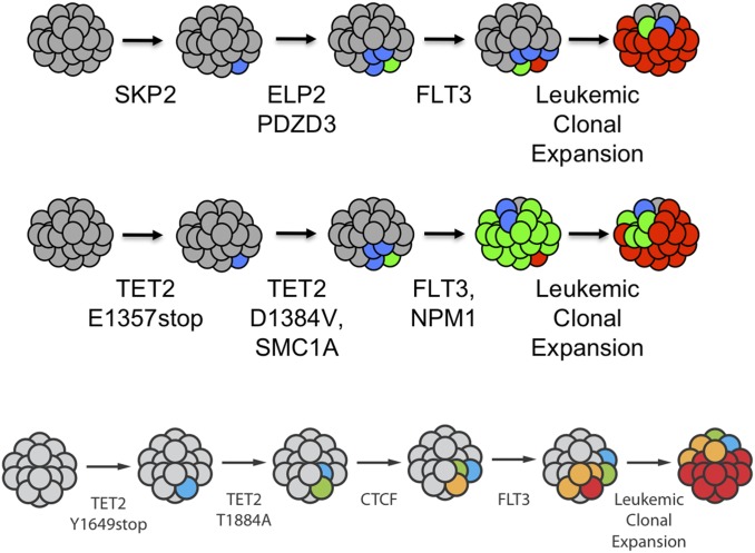
It is obvious that natural selection operates at the level of individuals and collections of individuals. Nearly two decades ago we showed that in multi-individual colonies of protochordate colonial tunicates sharing a blood circulation, there exists an exchange of somatic stem cells and germline stem cells, resulting in somatic chimeras and stem cell competitions for gonadal niches. Stem cells are unlike other cells in the body in that they alone self-renew, so that they form clones that are perpetuated for the life of the organism. Stem cell competitions have allowed the emergence of competitive somatic and germline stem cell clones. Highly successful germline stem cells usually outcompete less successful competitors both in the gonads of the genotype partner from which they arise and in the gonads of the natural parabiotic partners. Therefore, natural selection also operates at the level of germline stem cell clones. In the colonial tunicate Botryllus schlosseri the formation of natural parabionts is prevented by a single-locus highly polymorphic histocompatibility gene called Botryllus histocompatibility factor. This limits germline stem cell predation to kin, as the locus has hundreds of alleles. We show that in mice germline stem cells compete for gonad niches, and in mice and humans, blood-forming stem cells also compete for bone marrow niches. We show that the clonal progression from blood-forming stem cells to acute leukemias by successive genetic and epigenetic events in blood stem cells also involves competition and selection between clones and propose that this is a general theme in cancer.

Keywords: Botryllus schlosseri; cancer; evolution; natural selection; stem cell competition.


Conflict of interest statement

The author declares no conflict of interest.

Figures

Fig. 1.
Fig. 1.
Life cycle of B. schlosseri. B. schlosseri reproduces both through sexual and asexual (budding) pathways, giving rise to virtually identical adult body plans. Upon settlement, the chordate tadpole, the product of sexual reproduction, metamorphoses into an invertebrate founder individual, an oozooid. This oozoid undergoes asexual reproduction through budding that proceeds through four stages (AD) to create a colony (Left) of genetically identical individuals (blastozooids, also known as zooids). This budding process continues weekly throughout the life of the colony producing multiple individuals (buds that grow into zooids). Each individual has anatomical features that include atrial and oral siphons as well as a simple tube-like heart, intestines, and a branchial sac. The individuals are connected via a network of blood vessels that are embedded within a gelatinous matrix (termed “tunic”) and terminate in finger-like protrusions (ampullae). Reproduced with permission from ref. .
Fig. 2.
Fig. 2.
Fusion/rejection between B. schlosseri colonies is governed by the BHF gene. Colonies that share an allele will fuse (Left), whereas colonies that do not will reject (Right). Rejection is an inflammatory response of blood cells in ampullae and results in a fibrotic scar that blocks vascular anastomoses. Major anatomical features are indicated (amp, ampullae, the colony’s blind-ended vasculature). Adapted with permission from ref. .
Fig. 3.
Fig. 3.
An autoradiograph and interpretative table that show the results of genotypic sampling of buds (B), sperm (S), and blood (BL) from a field chimera of B. schlosseri in which we observed germ cell or somatic cell parasitism (G/SCP, the phenomenon whereby the stem cells of one chimeric partner take over the tissue of the other chimeric partner in a colony). The tissues were typed using microsatellite locus BS811 (6). Based on the spatial pattern of genotypes within the somatic tissues of the chimera at the time of collection, it is presumed that the initial genotypes for the two colonies were AA (colony 1) and AB (colony 2). After fusion, both the somatic and gametic tissues of colony AA appear to remain unchanged. In contrast, whereas the somatic tissues of colony AB retained their original genotype, its gametic tissues were almost completely replaced by tissues having genotype AA. This replacement is interpreted as an example of G/SCP. Reproduced with permission from ref. .
Fig. 4.
Fig. 4.
Hierarchies of germline stem cell and somatic stem cell competition. Three genetically distinct of colonies (strains F, B, and G) with shared BHF alleles were allowed to fuse in all pair-wise combinations and also all together to form chimeric colonies. (n = 3, Fusion Partners). After 3 mo, the sperm (Middle) and somatic tissue (Right) were analyzed to identify which strains contributed to each tissue. In most cases, a single germline “winner” outcompeted the germline loser, and the outcomes of this competition to dominate spermatogenesis followed a hierarchical relationship. The allelic markers shown here identify the majority of cells in the testis. Both testis and ovaries from each colony gave rise to gametes that were tested in crosses. If the germline allelic marker in the testis assay won, the sperm and eggs from that colony came from the winner (F dominated over G, which dominated over B) (8).
Fig. 5.
Fig. 5.
Model of clonal frequencies of germline (or somatic) stem cells in an individual. (Left) Equally competitive stem cells coexist. (Center) Supercompetitor stem cells expand at the expense of normal stem cells, but the supercompetitor stem cells + normal stem cells do not exceed the stem cell frequency for that type of stem cell. (Right) The heritable changes in a stem cell lineage allow the clone to expand dramatically and beyond the frequency limitation of the previous part. Here the stem cell resembles a cancer stem cell.
Fig. 6.
Fig. 6.
Many adjacent seminiferous tubules within a testis derive from a single germline stem cell. To define how many germline stem cells ultimately contribute to the seminiferous tubules of the adult testes, a clonal analysis of stem cell progeny was performed. Tetrachimeric mice harbor cells that are permanently labeled with one of four marks: green (eGFP), blue (eCFP), red (mRFP1), or fluorescently uncolored. The dissected and untangled seminiferous tubules from these tetrachimeras show that each tubule is a single color and that multiple adjacent tubules are all of the same color (i.e., derived from a single germline stem cell). Images are representative of 32 chimeric testes analyzed; examples are of (A) a testes with only blue or red tubules; (B and C) a testes with only blue or green tubules; and (D) a testes with only red and green tubules. Reproduced with permission from ref. .
Fig. 7.
Fig. 7.
Our model of two-step oligoclonal development of testis germ cells. Male germline starts from four cells. They proliferate, migrate, and split to bilateral genital ridges. However, germ cells actually contributing adult spermatogenesis during the reproductive period originate from a small number of a second founder population that initially seed onto genital ridges. The rest of the germ cells (deciduous germ cells) are removed, most likely by apoptosis, before the reproductive period. Reproduced with permission from ref. .
Fig. 8.
Fig. 8.
Model of leukemic progression. In this model, HSCs serve as the reservoir for the accumulation of the genetic and epigenetic events that eventually lead to blast crisis and leukemia. Stem cell self-renewal and differentiation enable heritable mutations acquired in the stem cell compartment to be propagated to both self-renewing progeny and downstream progenitors over the lifetime of the organism. Stem cells with heritable lesions act as substrates for additional hits, which in turn can promote selection of preleukemic clones if lesions imparting a growth or survival advantage are acquired. In this model, seven events are listed as the full complement of events required for the progression of preleukemic clones to leukemia, but the actual number of events may vary depending upon the type of cancer and the nature of the lesions involved as it is possible that multiple oncogenic properties could be conferred upon a preleukemic clone through the acquisition of a single hit. Although the mutagenic events accrue in stem cells, the eventual emergence of leukemic clones occurs at the stage of progenitor cells downstream of HSCs that acquire the capacity for unlimited self-renewal. In CML, this can be the granulocyte/macrophage progenitor, whereas, in acute myeloid leukemia, the leukemic clone can emerge at the level of a multipotent progenitor or at a stage further downstream depending upon the nature of the oncogenic lesions involved. Reproduced with permission from ref. .
Fig. 9.
Fig. 9.
Model of aging HSCs. In young humans and mice, the HSC pool is dominated by cells that have a balance of myeloid and lymphoid lineage fates (orange circles). In older humans and mice, HSCs with a bias toward myeloid fates dominate (blue circles). Adapted from data presented in ref .
Fig. 10.
Fig. 10.
Single-cell analysis identifies sequential acquisition of mutations in preleukemic HSCs. The schematic in each row shows models for the proposed clonal evolution of acute myeloid leukemia in each of three different patients. Adapted with permission from ref. .

References

    1. Weissman IL. Stem cells: Units of development, units of regeneration, and units in evolution. Cell. 2000;100(1):157–168. - PubMed
    1. Laird DJ, Weissman IL. Telomerase maintained in self-renewing tissues during serial regeneration of the urochordate Botryllus schlosseri. Dev Biol. 2004;273(2):185–194. - PubMed
    1. Scofield VL, Schlumpberger JM, West LA, Weissman IL. Protochordate allorecognition is controlled by a MHC-like gene system. Nature. 1982;295(5849):499–502. - PubMed
    1. Voskoboynik A, et al. Identification of a colonial chordate histocompatibility gene. Science. 2013;341(6144):384–387. - PMC - PubMed
    1. Voskoboynik A, et al. The genome sequence of the colonial chordate, Botryllus schlosseri. eLife. 2013;2:e00569. - PMC - PubMed

Publication types