

Detection of tumor-derived DNA in cerebrospinal fluid of patients with primary tumors of the brain and spinal cord
- PMID: 26195750
- PMCID: PMC4534284
- DOI: 10.1073/pnas.1511694112
Detection of tumor-derived DNA in cerebrospinal fluid of patients with primary tumors of the brain and spinal cord
Abstract
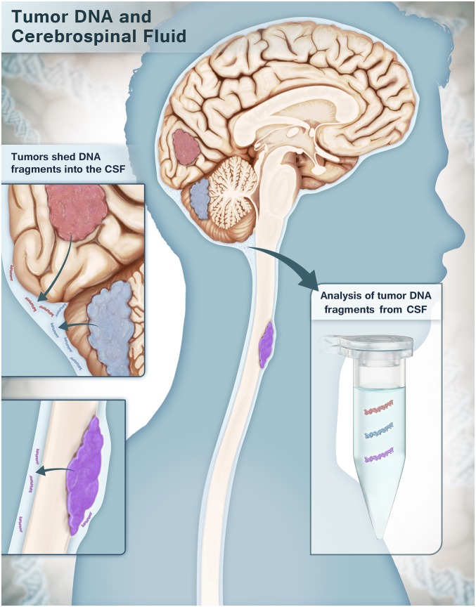
Cell-free DNA shed by cancer cells has been shown to be a rich source of putative tumor-specific biomarkers. Because cell-free DNA from brain and spinal cord tumors cannot usually be detected in the blood, we studied whether the cerebrospinal fluid (CSF) that bathes the CNS is enriched for tumor DNA, here termed CSF-tDNA. We analyzed 35 primary CNS malignancies and found at least one mutation in each tumor using targeted or genome-wide sequencing. Using these patient-specific mutations as biomarkers, we identified detectable levels of CSF-tDNA in 74% [95% confidence interval (95% CI) = 57-88%] of cases. All medulloblastomas, ependymomas, and high-grade gliomas that abutted a CSF space were detectable (100% of 21 cases; 95% CI = 88-100%), whereas no CSF-tDNA was detected in patients whose tumors were not directly adjacent to a CSF reservoir (P < 0.0001, Fisher's exact test). These results suggest that CSF-tDNA could be useful for the management of patients with primary tumors of the brain or spinal cord.
Keywords: CNS tumors; CSF-tDNA; biomarker.
Conflict of interest statement
The authors declare no conflict of interest.
Figures
References
Publication types
MeSH terms
Substances
Grants and funding
LinkOut - more resources
Full Text Sources
Other Literature Sources
Medical
