

Quantification of Multifidus Atrophy and Fatty Infiltration Following a Minimally Invasive Microdiscectomy
- PMID: 26196032
- PMCID: PMC4505388
- DOI: 10.14444/2025
Quantification of Multifidus Atrophy and Fatty Infiltration Following a Minimally Invasive Microdiscectomy
Abstract
Background: Multifidus muscle degeneration and atrophy have been demonstrated following traditional open spine surgery. The purpose of this study was to quantify multifidus muscle atrophy and fatty infiltration following a 1-level minimally invasive (MIS) lumbar discectomy.
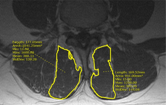
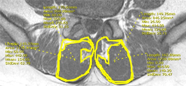
Methods: Magnetic resonance imaging (MRI) of 24 patients who underwent a primary 1-level MIS microdiscectomy were reviewed. Demographics, operative levels, and time from surgery to imaging were assessed. Total and lean cross-sectional areas (CSA), T1-signal intensity ratio between the multifidus and psoas muscles, and lean-to-total CSA ratio were measured. Pre- and postoperative values were compared within each patient utilizing paired sample T-tests.
Results: The mean age was 47.8±14.2 years. MRI was obtained 182.5±194.4 days following index surgery. On the ipsilateral side, total CSA decreased at the index level (-4.9%) and the lean CSA decreased at the index (-6.2%), inferior pedicle (-13.0%), and inferior disc levels (-18.6%). On the contralateral side, no significant decreases in total or lean CSA were demonstrated. T1-signal intensity ratios increased at all levels, but the differences were not statistically significant. The lean-to-total CSA ratio was decreased at the superior disc (-5.2%), inferior pedicle (-8.4%), and inferior disc levels (-17.2%) on the ipsilateral side and at the contralateral inferior disc level (-5.3%).
Conclusions: Primary 1-level MIS discectomy results in minimal short-term atrophy and fatty infiltration of the multifidus at the index level. Total CSA atrophy was mainly confined to the ipsilateral side at the index level. Lean CSA atrophy was observed mainly at and below the index level on the ipsilateral side. Fatty infiltration, as measured by the lean-to-total CSA ratio, ranged 1.2-17.2% on the ipsilateral and 0-5.3% on the contralateral side with greater fat content demonstrated caudally to the surgical level.
Clinical relevance: Overall, the majority of the multifidus muscle appears to be radiographically preserved following an MIS lumbar discectomy.
Keywords: MRI multifidus; Multifidus atrophy; T1 signal intensity; minimally invasive multifidus; multifidus fat infiltration; paraspinal atrophy.
Figures
References
-
- Cholewicki J, Panjabi MM, Khachatryan A. Stabilizing function of trunk flexor-extensor muscles around a neutral spine posture. Spine. 1997 Oct 1;22(19):2207–2212. - PubMed
-
- Macintosh JE, Bogduk N. 1987 Volvo award in basic science. The morphology of the lumbar erector spinae. Spine. 1987 Sep;12(7):658–668. - PubMed
-
- Macintosh JE, Bogduk N. The attachments of the lumbar erector spinae. Spine. 1991 Jul;16(7):783–792. - PubMed
-
- Freeman MD, Woodham MA, Woodham AW. The role of the lumbar multifidus in chronic low back pain: a review. PM & R: the journal of injury, function, and rehabilitation. 2010 Feb;2(2):142–146. quiz 141 pfollowing 167. - PubMed
LinkOut - more resources
Full Text Sources
Other Literature Sources