. 2015 Jul 21;10(7):e0133321.
doi: 10.1371/journal.pone.0133321. eCollection 2015.

Eukaryotic rRNA Modification by Yeast 5-Methylcytosine-Methyltransferases and Human Proliferation-Associated Antigen p120

Affiliations

Eukaryotic rRNA Modification by Yeast 5-Methylcytosine-Methyltransferases and Human Proliferation-Associated Antigen p120

Gabrielle Bourgeois et al. PLoS One. .

Abstract

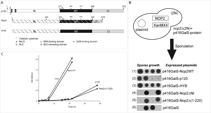
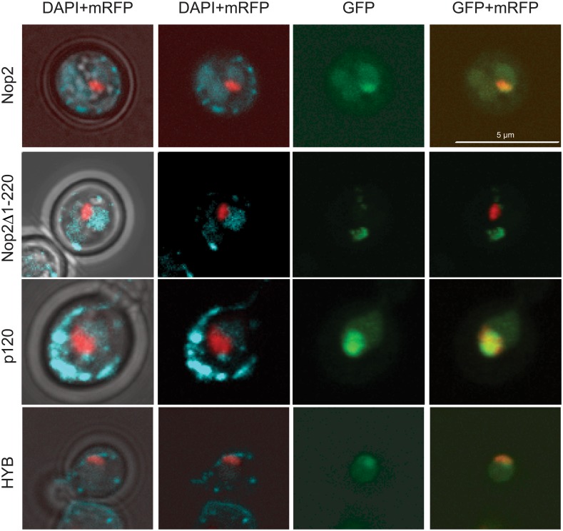
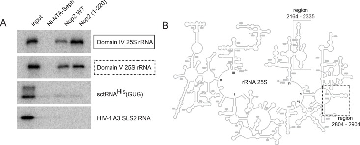
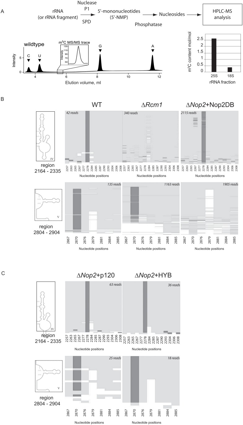
Modified nucleotide 5-methylcytosine (m5C) is frequently present in various eukaryotic RNAs, including tRNAs, rRNAs and in other non-coding RNAs, as well as in mRNAs. RNA:m5C-methyltranferases (MTases) Nop2 from S. cerevisiae and human proliferation-associated nucleolar antigen p120 are both members of a protein family called Nop2/NSUN/NOL1. Protein p120 is well-known as a tumor marker which is over-expressed in various cancer tissues. Using a combination of RNA bisulfite sequencing and HPLC-MS/MS analysis, we demonstrated here that p120 displays an RNA:m5C- MTase activity, which restores m5C formation at position 2870 in domain V of 25S rRNA in a nop2Δ yeast strain. We also confirm that yeast proteins Nop2p and Rcm1p catalyze the formation of m5C in domains V and IV, respectively. In addition, we do not find any evidence of m5C residues in yeast 18S rRNA. We also performed functional complementation of Nop2-deficient yeasts by human p120 and studied the importance of different sequence and structural domains of Nop2 and p120 for yeast growth and m5C-MTase activity. Chimeric protein formed by Nop2 and p120 fragments revealed the importance of Nop2 N-terminal domain for correct protein localization and its cellular function. We also validated that the presence of Nop2, rather than the m5C modification in rRNA itself, is required for pre-rRNA processing. Our results corroborate that Nop2 belongs to the large family of pre-ribosomal proteins and possesses two related functions in pre-rRNA processing: as an essential factor for cleavages and m5C:RNA:modification. These results support the notion of quality control during ribosome synthesis by such modification enzymes.


Conflict of interest statement

Competing Interests: The authors have declared that no competing interests exist.

Figures

Fig 1
Fig 1. Protein domain structures of human p120 and S. cerevisiae Nop2 and conservation of the putative RNA:m5C-MTase active site.
(A) Global alignment of human p120, yeast Nop2 and HYB protein. Proteins are represented to scale and domains present in p120 and Nop2 as well as their positions are indicated (NTD: N-terminal domain, MTD: RNA:MTase catalytic domain, CTD: C-terminal domain). The positions of catalytic cysteine residues in the MTD are indicated by asterisks. Specific sequence motifs, identified in p120 and Nop2, are indicated by letters: a: p120 Nucleolar Localization Signal (NoLS), b: p120 Nuclear Localization Signal (NLS), c: RNA-binding domain, d: B23-interacting domain, e: AdoMet (SAM) binding domain. In HYB protein, p120 structural domains are indicated as grey and black boxes and the corresponding Nop2 domains are indicated as hatched white rectangle. (B) Preparation of haploid Nop2Δ strains by sporulation and tetrad dissection. Growth of individual spores expressing different variants is shown at the bottom. (C) Growth of viable complemented haploid strains in liquid YPG medium. Strains were inoculated at 0.05−0.1 units OD600 and grown for a maximum of 40 hours on a shaker at 30°C.
Fig 2
Fig 2. Subcellular localization of WT Nop2, p120, Nop2Δ1–220 and chimeric HYB protein.
GFP-tagged variants were expressed in the presence of mRFP-Nop56 [46] and the endogenous Nop2 in yeast. Cells were DAPI stained and imaged in vivo. Columns from left to right: (1) merge of brightfield, DAPI and mRFP-Nop56 channels to establish the borders of the cells, the nuclei and nucleoli, respectively; (2) DAPI and mRFP-Nop56 only; (3) GFP-tagged protein as indicated to the left of the rows; and (4) merge between GFP-tagged protein and mRFP-Nop56 signals to highlight the possible co-localization of the two signals. The scale bar is 5 μm. Quantification of nuclear and nucleolar signals is given in the Table A in S1 Text.
Fig 3
Fig 3. Specific rRNA binding by recombinant full-length Nop2 and its N-terminal fragment.
(A) His6-tagged recombinant protein was immobilized on Ni-NTA Sepharose beads and incubated with radiolabeled transcripts of Domains IV and V of S. cerevisiae 25S rRNA (fragments spanning nucleotides 2164−2335 and 2804−2904, respectively), RNAs derived from S. cerevisiae tRNAHis(GUG), HIV-1 derived RNA covering stem-loop structure 2 (SLS2) of the A3 splice site [47] and also transcripts corresponding to yeast ITS1 and ITS2 rRNA sequences. After extensive washing to remove unbound RNA, the retained fraction was directly loaded onto SDS-PAGE and analyzed by autoradiography. 5% of the input was loaded in the first lane on the left. The second lane shows the RNA bound without immobilized protein. (B) Schematic representation of 25S yeast rRNA: the well-defined structural domains are numbered from I to VI, and the regions corresponding to tested in vitro transcripts are boxed (solid box–Domain IV, dashed box–Domain V).
Fig 4
Fig 4
(A) Analysis of m5C presence in full length yeast rRNA. Schematic representation of the procedure is given on the top and typical separation profile for nucleosides at the bottom. Quantification of m5C residues in full-length 25S and 18S rRNAs is presented by the histogram. (B) Results of bisulfite sequencing of Domains IV and V of 25S rRNA extracted from different deleted and complemented yeast strains. Dark gray shade indicate bisulfite resistant non-deaminated m5C residue, light shade represents deamination events C → U. Numbering of all C residues present in the domain is given at the bottom of each panel. Number of reads mapped to a given region is indicated on the top. (C) Bisulfite sequencing of 25S rRNA extracted from yeast strains expressing human p120 and HYB proteins. The legend is the same as for panel B.

Similar articles

Cited by

References

    1. Motorin Y, Helm M. RNA nucleotide methylation. Wiley Interdiscip Rev RNA 2011;2(5):611–31. 10.1002/wrna.79 - DOI - PubMed
    1. Salozhin SV, Prokhorchuk EB, Georgiev GP. Methylation of DNA—one of the major epigenetic markers. Biochemistry (Mosc) 2005;70(5):525–32. - PubMed
    1. Smith JE, Cooperman BS, Mitchell P. Methylation sites in Escherichia coli ribosomal RNA: localization and identification of four new sites of methylation in 23S rRNA. Biochemistry 1992;31(44):10825–34. - PubMed
    1. Veldman GM, Klootwijk J, de Regt VC, Planta RJ, Branlant C, Krol A, et al. The primary and secondary structure of yeast 26S rRNA. Nucleic Acids Res 1981;9(24):6935–52. - PMC - PubMed
    1. Brand RC, Klootwijk J, Van Steenbergen TJ, De Kok AJ, Planta RJ. Secondary methylation of yeast ribosomal precursor RNA. Eur J Biochem 1977;75(1):311–8. - PubMed

Publication types

MeSH terms