Review
. 2015 Nov:49:17-45.
doi: 10.1016/j.preteyeres.2015.07.002. Epub 2015 Jul 18.

Progress in corneal wound healing

Affiliations
Review

Progress in corneal wound healing

Alexander V Ljubimov et al. Prog Retin Eye Res. 2015 Nov.

Abstract

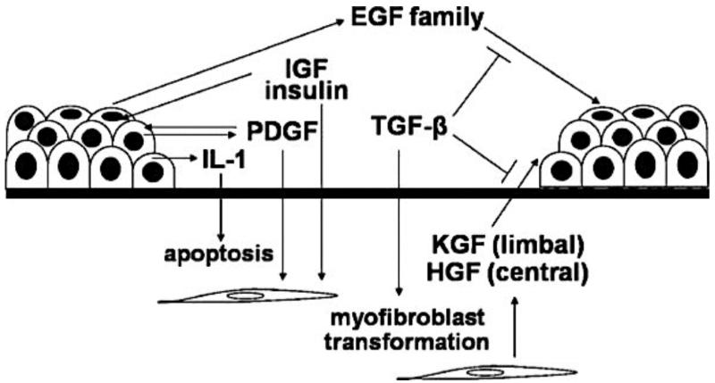
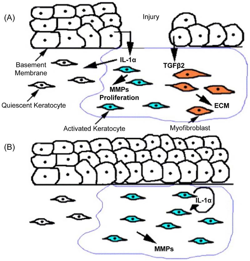
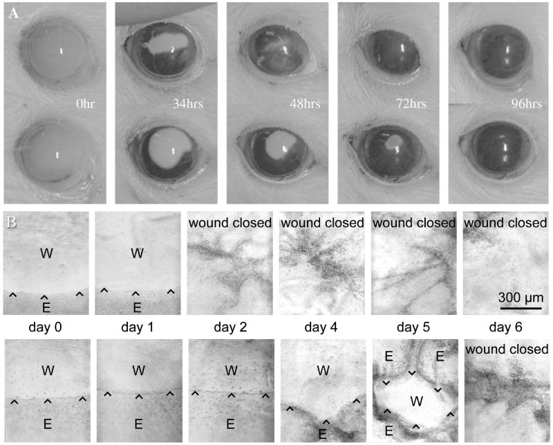
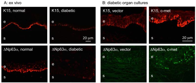
Corneal wound healing is a complex process involving cell death, migration, proliferation, differentiation, and extracellular matrix remodeling. Many similarities are observed in the healing processes of corneal epithelial, stromal and endothelial cells, as well as cell-specific differences. Corneal epithelial healing largely depends on limbal stem cells and remodeling of the basement membrane. During stromal healing, keratocytes get transformed to motile and contractile myofibroblasts largely due to activation of transforming growth factor-β (TGF-β) system. Endothelial cells heal mostly by migration and spreading, with cell proliferation playing a secondary role. In the last decade, many aspects of wound healing process in different parts of the cornea have been elucidated, and some new therapeutic approaches have emerged. The concept of limbal stem cells received rigorous experimental corroboration, with new markers uncovered and new treatment options including gene and microRNA therapy tested in experimental systems. Transplantation of limbal stem cell-enriched cultures for efficient re-epithelialization in stem cell deficiency and corneal injuries has become reality in clinical setting. Mediators and course of events during stromal healing have been detailed, and new treatment regimens including gene (decorin) and stem cell therapy for excessive healing have been designed. This is a very important advance given the popularity of various refractive surgeries entailing stromal wound healing. Successful surgical ways of replacing the diseased endothelium have been clinically tested, and new approaches to accelerate endothelial healing and suppress endothelial-mesenchymal transformation have been proposed including Rho kinase (ROCK) inhibitor eye drops and gene therapy to activate TGF-β inhibitor SMAD7. Promising new technologies with potential for corneal wound healing manipulation including microRNA, induced pluripotent stem cells to generate corneal epithelium, and nanocarriers for corneal drug delivery are discussed. Attention is also paid to problems in wound healing understanding and treatment, such as lack of specific epithelial stem cell markers, reliable identification of stem cells, efficient prevention of haze and stromal scar formation, lack of data on wound regulating microRNAs in keratocytes and endothelial cells, as well as virtual lack of targeted systems for drug and gene delivery to select corneal cells.

Keywords: Corneal endothelium; Corneal epithelium; Gene therapy; Keratocyte; Stem cell; Wound healing.


Figures

Figure 1
Figure 1
Growth factors and epithelial wound healing. A multitude of growth factors and cytokines is released following an epithelial injury in the cornea. These factors play essential roles in epithelial–stromal interaction and in the successful healing of a wound. KGF and HGF are believed to be produced by keratocytes to influence epithelial behaviors, while IL-1 and PDGF may be master mediators secreted by the epithelium to modulate stromal response to injury. Others such as the EGF family, IGF, and TGF-β regulate both the epithelium and stromal cell transformation to myofibroblasts, and the cross-talk among various growth factors determines the outcome of an epithelial wound. Reproduced with permission from Yu et al., 2010b.
Figure 2
Figure 2
Cellular interactions during corneal repair. (A) Upon corneal epithelial injury, IL-1α is released from the injured epithelium into the stroma. IL-1α induces some of the underlying stromal keratocytes to undergo cell death, while others are induced to proliferate, secrete MMPs, and transition from a quiescent to an activated phenotype. Due to the absence of a basement membrane, corneal epithelial cells also secrete TGF-β2 into the underlying stroma inducing a subpopulation of keratocytes to undergo transformation into myofibroblasts that secrete ECM. (B) The return of the basement membrane inhibits the release of TGF-β2 into the stroma and the myofibroblast phenotype is no longer observed. The activated keratocytes continue to secrete autocrine IL-1α and remodel the ECM. Reproduced with permission from West-Mays and Dwivedi, 2006.
Figure 3
Figure 3
Representative wound healing dynamics of normal and diabetic corneas. A, healing of rat epithelial scrape wounds. Upper panel, normal rat eye; lower panel, Goto-Kakizaki diabetic rat eye (DM2 model). Healing of diabetic cornea is clearly delayed. Reproduced with permission from Chikama et al., 2007. B, healing of n-heptanol induced epithelial wounds in organ-cultured human corneas. Upper panel, normal cornea; lower panel, diabetic cornea. Healing of diabetic cornea is significantly delayed. Wound edges are marked by arrowheads. W, wound; E, epithelium. Reproduced with permission from Kabosova et al., 2003.
Figure 4
Figure 4
Gene therapy-mediated upregulation of stem cell marker expression in diabetic corneas. A. Putative LESC marker expression patterns in normal and diabetic ex vivo limbus. Note a dramatic decrease in staining intensity and the number of positive basal epithelial cells for K15 and ΔNp63α in the diabetic limbus. B. Increased putative LESC marker expression in the diabetic limbus in organ culture upon c-met overexpression. c-Met gene transduction led to elevated and similar to normal expression of K15 and ΔNp63α in the limbus of organ-cultured diabetic corneas. e, epithelium, s, stroma. Bars = 20 μm. Reproduced from Saghizadeh et al., 2011.
Figure 5
Figure 5
Increased expression of diabetic markers upon proteinase gene silencing in organ-cultured diabetic corneas. Left, limbal BM component laminin γ3 chain; a markedly increased staining and continuity is seen after shRNA silencing of MMP-10 (M10) and cathepsin F (CF) expression. Right, similar results obtained for a diabetes-downregulated marker α3β1 integrin. Bar = 30 μm. Reproduced from Saghizadeh et al., 2013b. © Association for Research in Vision and Ophthalmology.
Figure 6
Figure 6
Wound healing in miR-146a inhibitor transfected human diabetic organ-cultured corneas. A. Transfection with miR-146a inhibitor enhanced wound healing compared to control transfected with labeled scrambled miR-Cy3. Transfected diabetic organ-cultured cornea with miR-146a inhibitor, upper row; with miR-Cy3-scrambled control, lower row. E, epithelium; W, wounded area. B. Quantitation of wound healing rates. The bar graph represents average ± SEM of pooled values (n=6) of days to heal. ** p<0.001 by paired two-tailed t test. Reproduced from Winkler et al., 2014.

References

    1. Aberdam E, Barak E, Rouleau M, de LaForest S, Berrih-Aknin S, Suter DM, Krause KH, Amit M, Itskovitz-Eldor J, Aberdam D. A pure population of ectodermal cells derived from human embryonic stem cells. Stem Cells. 2008;26:440–444. - PubMed
    1. Abrams GA, Schaus SS, Goodman SL, Nealey PF, Murphy CJ. Nanoscale topography of the corneal epithelial basement membrane and Descemet’s membrane of the human. Cornea. 2000;19:57–64. - PubMed
    1. Adijanto J, Naash MI. Nanoparticle-based technologies for retinal gene therapy. Eur. J. Pharm. Biopharm. 2015;12:S0939–6411. - PMC - PubMed
    1. Ahmad S, Kolli S, Lako M, Figueiredo F, Daniels JT. Stem cell therapies for ocular surface disease. Drug Discov. Today. 2010;15:306–313. - PubMed
    1. Alio JL, Pastor S, Ruiz-Colecha J, Rodriguez A, Artola A. Treatment of ocular surface syndrome after LASIK with autologous platelet-rich plasma. J. Refract. Surg. 2007;23:617–619. - PubMed

Publication types

MeSH terms