. 2015 Jul 21;11(7):e1005051.
doi: 10.1371/journal.ppat.1005051. eCollection 2015 Jul.

K-bZIP Mediated SUMO-2/3 Specific Modification on the KSHV Genome Negatively Regulates Lytic Gene Expression and Viral Reactivation

Affiliations

K-bZIP Mediated SUMO-2/3 Specific Modification on the KSHV Genome Negatively Regulates Lytic Gene Expression and Viral Reactivation

Wan-Shan Yang et al. PLoS Pathog. .

Abstract

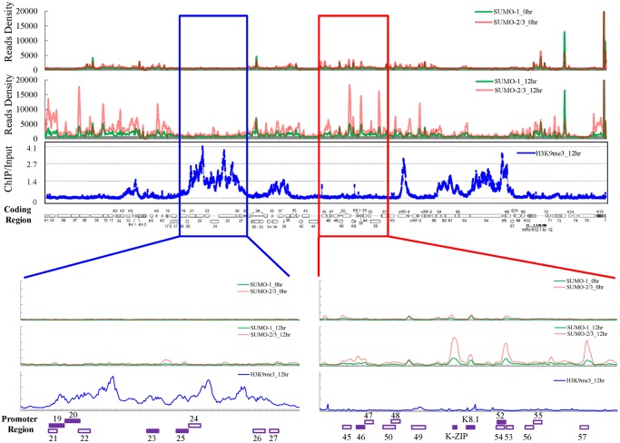
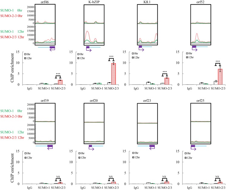
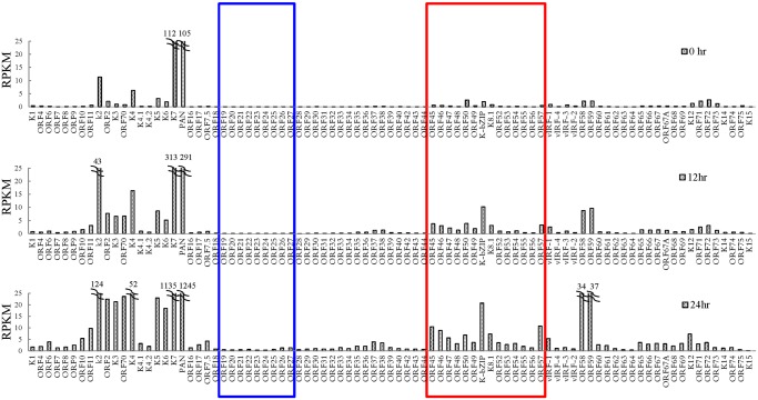
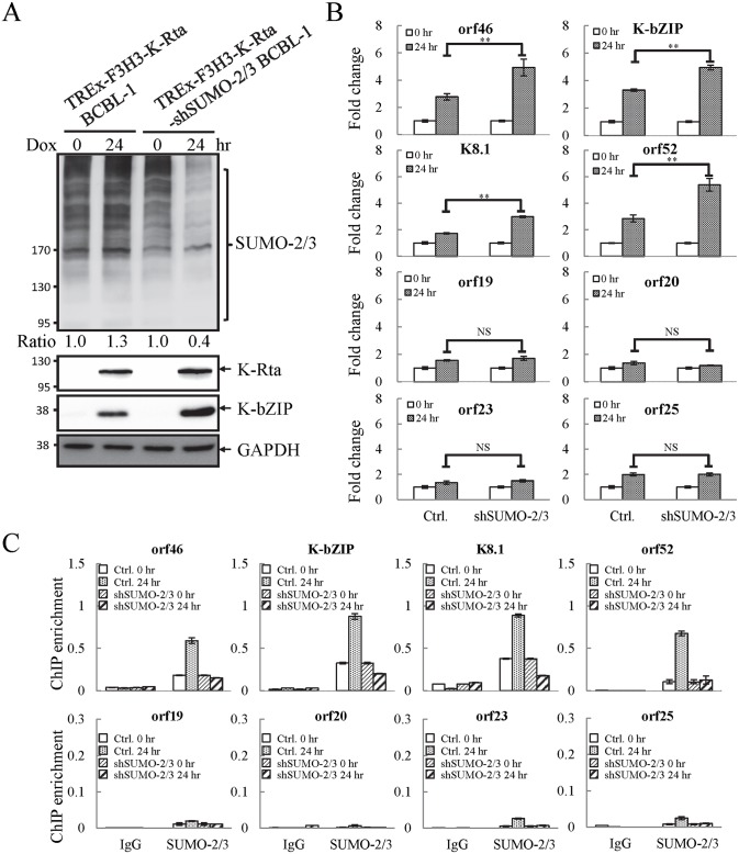
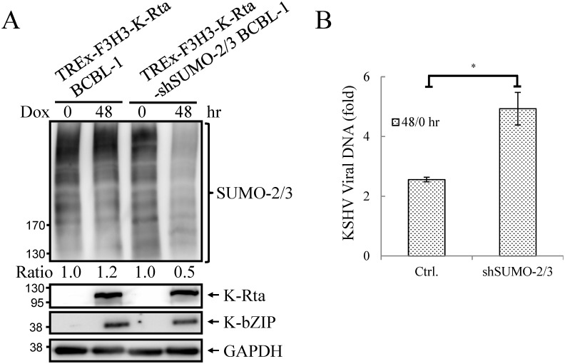
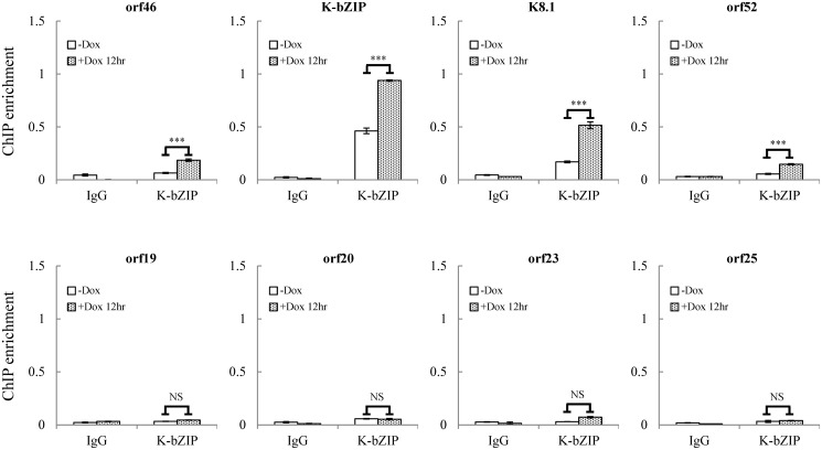
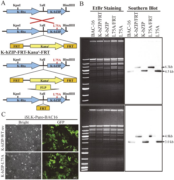
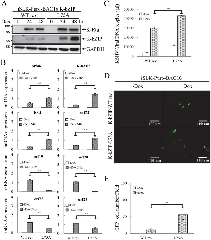
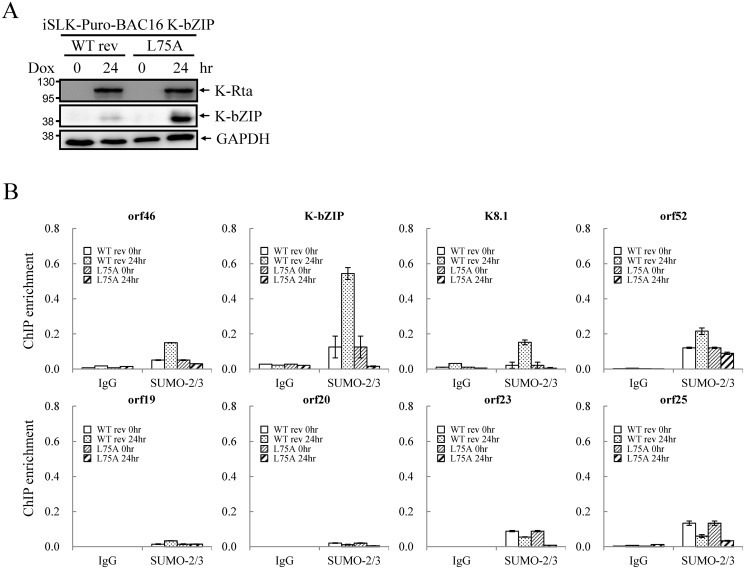
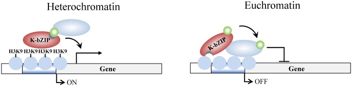
SUMOylation is associated with epigenetic regulation of chromatin structure and transcription. Epigenetic modifications of herpesviral genomes accompany the transcriptional switch of latent and lytic genes during the virus life cycle. Here, we report a genome-wide comparison of SUMO paralog modification on the KSHV genome. Using chromatin immunoprecipitation in conjunction with high-throughput sequencing, our study revealed highly distinct landscape changes of SUMO paralog genomic modifications associated with KSHV reactivation. A rapid and widespread deposition of SUMO-2/3, compared with SUMO-1, modification across the KSHV genome upon reactivation was observed. Interestingly, SUMO-2/3 enrichment was inversely correlated with H3K9me3 mark after reactivation, indicating that SUMO-2/3 may be responsible for regulating the expression of viral genes located in low heterochromatin regions during viral reactivation. RNA-sequencing analysis showed that the SUMO-2/3 enrichment pattern positively correlated with KSHV gene expression profiles. Activation of KSHV lytic genes located in regions with high SUMO-2/3 enrichment was enhanced by SUMO-2/3 knockdown. These findings suggest that SUMO-2/3 viral chromatin modification contributes to the diminution of viral gene expression during reactivation. Our previous study identified a SUMO-2/3-specific viral E3 ligase, K-bZIP, suggesting a potential role of this enzyme in regulating SUMO-2/3 enrichment and viral gene repression. Consistent with this prediction, higher K-bZIP binding on SUMO-2/3 enrichment region during reactivation was observed. Moreover, a K-bZIP SUMO E3 ligase dead mutant, K-bZIP-L75A, in the viral context, showed no SUMO-2/3 enrichment on viral chromatin and higher expression of viral genes located in SUMO-2/3 enriched regions during reactivation. Importantly, virus production significantly increased in both SUMO-2/3 knockdown and KSHV K-bZIP-L75A mutant cells. These results indicate that SUMO-2/3 modification of viral chromatin may function to counteract KSHV reactivation. As induction of herpesvirus reactivation may activate cellular antiviral regimes, our results suggest that development of viral SUMO E3 ligase specific inhibitors may be an avenue for anti-virus therapy.


Conflict of interest statement

The authors have declared that no competing interests exist.

Figures

Fig 1
Fig 1. Global mapping of SUMO paralogs on latent and lytic KSHV genomes and of H3K9me3 on the KSHV genome during lytic reactivation.
ChIP-seq for SUMO paralogs was performed using chromatin prepared from non-induced (0 hour) and 0.2 μg/ml Dox-treated (12 hours) TREx-F3H3-K-Rta BCBL-1 cells. The ChIP-seq result of the KSHV genome was normalized with total reads from the human genome. ChIP-on-chip for H3K9me3 was performed using chromatin prepared from 0.2 μg/ml Dox-treated cells as described above. The rectangular areas are regions that comprise high SUMO-2/3 (red) or high H3K9me3 (blue) levels after KSHV reactivation (upper panel) and are further magnified on the lower portion of the figure. Promoters of viral genes in these regions are marked in bottom. Solid squares represent genes used for further analysis (lower panel).
Fig 2
Fig 2. Deposition of SUMO-2/3 modification on selected KSHV promoters representing high SUMO-2/3 enrichment or high H3K9me3 mark regions during reactivation.
ChIP was performed as described in Fig 1 using anti-SUMO-1 or anti-SUMO-2/3 antibodies. SUMO-1 and SUMO-2/3 binding to orf46, K-bZIP, K8.1, and orf52 promoters in SUMO-2/3 enrichment region and orf19, orf20, orf23, and orf25 promoters in H3K9me3-rich region were analyzed by real-time qPCR. An enlarged view of the corresponding ChIP-seq mapping presented in Fig 1 is shown above each ChIP-qPCR plot. Gene promoters are solid boxes and the direction of transcription is shown by arrows. Rabbit IgG was used as negative antibody control and enrichment is not visible in the ChIP-qPCR plots. ***; P<0.001. NS; non-significant.
Fig 3
Fig 3. Transcriptome profile of KSHV during viral reactivation.
Total RNA samples isolated from non-induced (0 hour) and 0.2 μg/ml Dox-induced (for 12 and 24 hours) TREx-F3H3-K-Rta BCBL-1 cells were subjected to RNA-seq. Expression of all KSHV genes are presented as reads per kilobase per million mapped (RPKM). The rectangular areas are regions that comprise high SUMO-2/3 (red) or high H3K9me3 (blue) levels after KSHV reactivation as in Fig 1.
Fig 4
Fig 4. Knockdown of SUMO-2/3 increases transactivation of KSHV lytic genes and reduces SUMO-2/3-specific modification located in the high SUMO-2/3 enrichment region during viral reactivation.
(A) TREx-F3H3-K-Rta BCBL-1 and TREx-F3H3-K-Rta-shSUMO-2/3 BCBL-1 cells were treated with Dox (0.2 μg/ml) for 24 hours. Total cell lysates (TCLs) were analyzed by immunoblotting using anti-SUMO-2/3, anti-K-Rta, and anti-K-bZIP antibodies. Anti-GAPDH antibody was used for loading control. Ratio for each cell line is the SUMO-2/3/GAPDH signal observed for Dox treatment at 0 (for shSUMO-2/3) and 24 hour using TREx-F3H3-K-Rta BCBL-1 cells at 0 hour set as 1.0. (B) Total RNA isolated from cells treated as described in (A) was reverse transcribed using oligo-d(T)18 primer. The expression level of four viral genes representing SUMO-2/3 enrichment region and four viral genes representing H3K9me3-rich region as described in Fig 1 were quantified by real-time qPCR. All reactions were run in triplicate and normalized against GAPDH. The fold change was computed by comparing induced values to their non-induced controls. ctrl.; control, TREx-F3H3-K-Rta BCBL-1. **; P<0.005. NS; non-significant. (C) ChIP was performed using chromatin prepared from cells treated as described in (A) using anti-SUMO-2/3 antibody. Rabbit IgG was used as negative antibody control and enrichment is not visible in some plots. SUMO-2/3 binding to SUMO-2/3 enrichment and H3K9me3-rich regions were analyzed by real-time qPCR using primer pairs specific for the KSHV loci as described in Fig 2.
Fig 5
Fig 5. Knockdown SUMO-2/3 enhances KSHV virus production.
(A) TCLs from non-induced (0 hour) and 0.2 μg/ml Dox-treated (48 hours) TREx-F3H3-K-Rta BCBL-1 and TREx-F3H3-K-Rta-shSUMO-2/3 BCBL-1 cells were analyzed by immunoblotting using antibodies as described in Fig 4A. Ratio for each cell line is the SUMO-2/3/GAPDH signal observed for Dox treatment at 0 (for shSUMO-2/3) and 48 hour using TREx-F3H3-K-Rta BCBL-1 cells at 0 hour set as 1.0. (B) Supernatants from TREx-F3H3-K-Rta BCBL-1 and SUMO-2/3 knockdown TREx-F3H3-K-Rta-shSUMO-2/3 BCBL-1 cells were collected and filtered at 0 and 48 hours after Dox (0.2 μg/ml) treatment. Virion-associated DNA was purified and KSHV DNA levels were determined by TaqMan qPCR. Mean ± SD. Fold was determined by KSHV DNA copy number/μl at 48 hour divided by KSHV DNA copy number/μl at 0 hour. ctrl.; control, TREx-F3H3-K-Rta BCBL-1. *; P<0.05.
Fig 6
Fig 6. K-bZIP binding on selected KSHV promoters in TREx-F3H3-K-Rta BCBL-1 cells.
ChIP was performed using chromatin prepared from cells treated as described in Fig 1 using anti-K-bZIP antibody. Rabbit IgG was used as negative antibody control. K-bZIP binding to orf46, K-bZIP, K8.1, and orf52 promoters in the SUMO-2/3 enriched region and orf19, orf20, orf23, and orf25 promoters in H3K9me3-rich region were analyzed by real-time qPCR.
Fig 7
Fig 7. Generation of K-bZIP-L75A mutated and wild-type revertant (WT rev) recombinant KSHV (rKSHV) in BAC16.
(A) Schematic representation of the recombineering procedures used for construction of K-bZIP-L75A mutants in BAC16. Recombination between the KSHV genomic locus (top) and the K-bZIP L75A targeting vector (bottom) is illustrated in the upper part of the figure. FLP recombinase-mediated removal of the targeting cassette is depicted in the lower portion. (B) Agarose gel and Southern blot analysis of recombineered BAC16 that was digested with KpnI (upper panel, left) or HindIII (lower panel, left) and probed with a radiolabeled K-bZIP DNA fragment (right). (C) Stable cell line of iSLK-Puro-BAC16 shows >90% GFP-positive cells. Phase (left) and FITC channel (right) images are shown.
Fig 8
Fig 8. The SUMO E3 ligase activity of K-bZIP is necessary to alleviate the increase in gene expression and virus production during reactivation.
(A) TCLs were collected from non-induced (0 hour) and 1 μg/ml Dox treated (for 24 and 48 hours) iSLK-Puro-BAC16 K-bZIP-WT rev and -L75A cell lines. Immunoblotting was used to confirm the induction of K-Rta and expression of K-bZIP. GAPDH was used as loading control. (B) Total RNA isolated from non-induced (0 hour) and 1 μg/ml Dox treated (24 hours) iSLK-Puro-BAC16 K-bZIP-WT rev and -L75A cells was reverse transcribed using oligo-d(T)18 primer and analyzed by real-time qPCR using specific primer pairs representing genes in SUMO-2/3 enrichment and H3K9me3-rich regions. All reactions were run in triplicate and normalized against GAPDH. (C) Supernatants from iSLK-Puro-BAC16 K-bZIP-WT rev and -L75A cells were collected at 0 and 48 hours after Dox (1 μg/ml) treatment and filtered. Virion-associated DNA was purified and KSHV DNA levels were determined by TaqMan qPCR. Mean ± SD. **; P<0.005. (D) 293T cells were infected with filtered supernatants harvested from iSLK-Puro-BAC16 K-bZIP-WT rev and -L75A cells treated with or without 1 μg/ml Dox for 72 hours. GFP positive cells were analyzed by fluorescence microscopy (FITC, 10X magnification) 48 hours after infection. (E) The GFP positive cells were quantified using the average from >20 microscopic fields. Mean ± SD. **; P<0.005.
Fig 9
Fig 9. SUMO E3 ligase activity of K-bZIP is essential for SUMO-2/3 modification of the viral genome during lytic reactivation.
(A) TCLs were collected from non-induced (0 hour) and 1 μg/ml Dox treated (for 24 hours) iSLK-Puro-BAC16 K-bZIP-WT rev and -L75A cell lines. Immunoblotting was used to confirm the induction of K-Rta and expression of K-bZIP. GAPDH was used as loading control. (B) ChIP was performed using chromatin prepared from cells treated as described in (A) using anti-SUMO-2/3 antibody. The SUMO-2/3 modification on selected promoters in SUMO-2/3 enrichment and H3K9me3-rich regions were analyzed by real-time qPCR. Rabbit IgG was used as negative antibody control and enrichment is not visible in some plots.
Fig 10
Fig 10. Model of K-bZIP regulation of viral gene expression in heterochromatin and euchromatin regions of the KSHV genome during reactivation.
In heterochromatin region, K-bZIP binds directly to H3K9me3 and modulates the viral gene activation by recruiting SUMOylated transcription regulators. In euchromatin region, K-bZIP binds directly or indirectly (through interacting with other transcription factors) to DNA and mediates SUMOylation-dependent transcription repression. Green circles, SUMO-2/3; blue ovals, unknown SUMOylated target; brown ovals, K-bZIP; blue circles, histones; H3K9, histone H3K9me3 mark.

References

    1. Garcia-Dominguez M, Reyes JC (2009) SUMO association with repressor complexes, emerging routes for transcriptional control. Biochim Biophys Acta 1789: 451–459. 10.1016/j.bbagrm.2009.07.001 - DOI - PubMed
    1. Gill G (2010) SUMO weighs in on polycomb-dependent gene repression. Mol Cell 38: 157–159. 10.1016/j.molcel.2010.04.006 - DOI - PubMed
    1. Maison C, Bailly D, Roche D, Montes de Oca R, Probst AV, et al. (2011) SUMOylation promotes de novo targeting of HP1alpha to pericentric heterochromatin. Nat Genet 43: 220–227. 10.1038/ng.765 - DOI - PubMed
    1. Cubenas-Potts C, Matunis MJ (2013) SUMO: a multifaceted modifier of chromatin structure and function. Dev Cell 24: 1–12. 10.1016/j.devcel.2012.11.020 - DOI - PMC - PubMed
    1. Rosonina E, Duncan SM, Manley JL (2010) SUMO functions in constitutive transcription and during activation of inducible genes in yeast. Genes Dev 24: 1242–1252. 10.1101/gad.1917910 - DOI - PMC - PubMed

Publication types

MeSH terms