Review
. 2016 May;142(5):895-904.
doi: 10.1007/s00432-015-2004-4. Epub 2015 Jul 22.

Lung cancer: developments, concepts, and specific aspects of the new WHO classification

Affiliations
Review

Lung cancer: developments, concepts, and specific aspects of the new WHO classification

Iver Petersen et al. J Cancer Res Clin Oncol. 2016 May.

Abstract

Introduction: Diagnostic methods and algorithms for the diagnosis of pulmonary neoplasms have considerably changed over the recent years. Based on large-scale molecular characterization studies and the development of targeted therapies, precise morphological, immunohistochemical, and molecular pathological tumor subtyping is now of utmost importance for evidence-based treatment decisions. Changes of diagnostic concepts initially referred to biopsies and cytology specimens but are now also transferred to resection specimens.

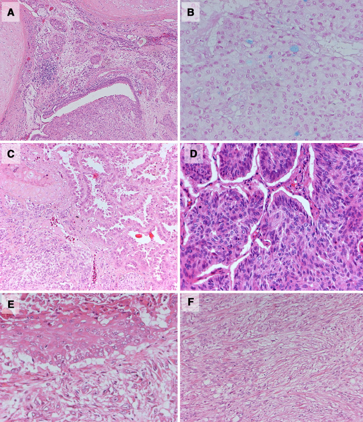
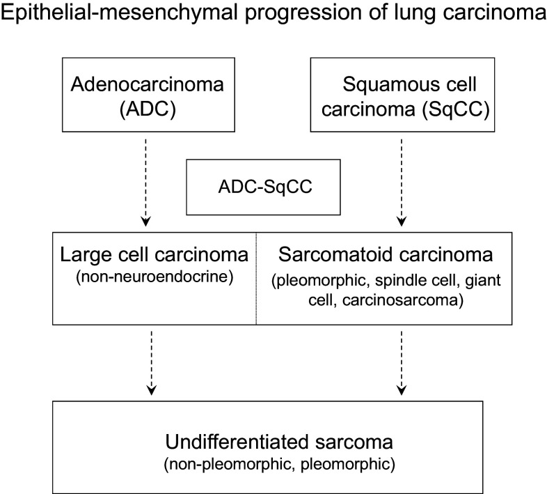
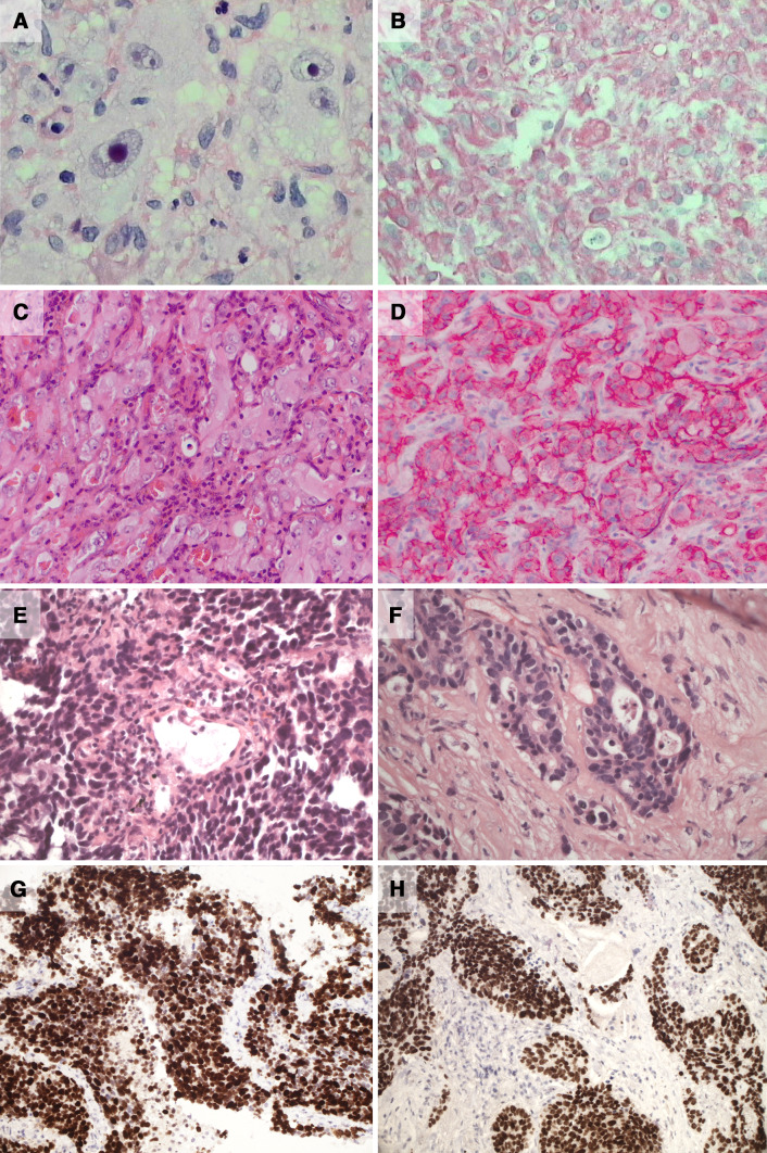
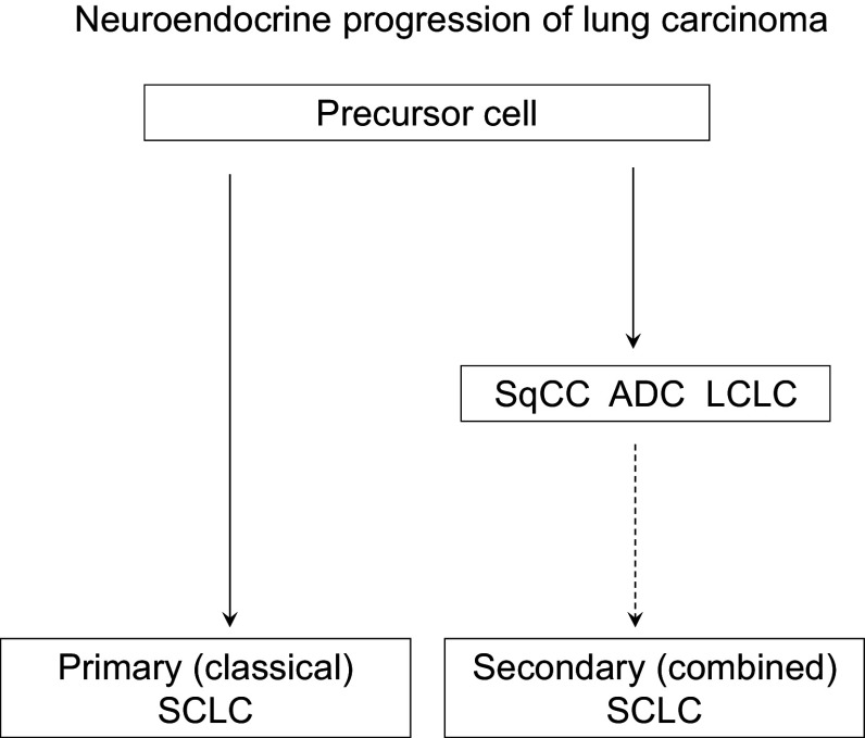
Methods: This review is focused on recent developments in morphological and immunohistochemical subtyping of pulmonary neoplasms and concepts of tumor progression. It also provides perspectives on relevant changes of diagnostic concepts within the context of the new WHO classification.

Conclusion: It becomes apparent that a three-step diagnostic concept based on morphology, immunohistochemistry, and molecular pathology is important to meet the requirements of an increasingly more complex, interdisciplinary care of lung cancer patients and to allow for reliable, clinically meaningful tumor diagnoses.

Keywords: Classification; Diagnosis; Immunohistochemistry; Lung cancer; Molecular pathology.


Conflict of interest statement

None.

Figures

Fig. 1
Fig. 1
a, b Lung adenocarcinoma with bronchial mucosa association, intraluminal growth, high degree of differentiation and little proliferation (a, HE). There is microfocal mucous formation (b, Alcian Blue) and signs of mucoepidermoid differentiation pointing to a tumor with characteristics of a mucoepidermoid salivary gland carcinoma. c Pulmonary adenocarcinoma with high degree of differentiation (see right upper part) and simultaneous hyperplasia of neuroendocrine cell at the border of a small bronchus (left lower part). d Basaloid squamous cell carcinoma of the lung with basaloid growth and focal retraction artefacts similar to a basal cell carcinoma of the skin. e, f Subtypes of sarcomatoid carcinomas of the lung. The pleomorphic carcinoma (e) is characterized by a combination of a non-small cell carcinoma, in this case of a squamous cell carcinoma (upper part) with a malignant sarcomatoid component similar to a spindle cell sarcoma (lower part). The distinction between a spindle cell carcinoma (f) and a sarcoma is only possible by the detection of a cytokeratin expression and/or an epithelial precursor lesion like a severe squamous dysplasia of a squamous cell carcinoma in situ within or at the border of the malignant spindle cell tumor
Fig. 2
Fig. 2
Epithelial–mesenchymal transition (EMT) of non-small cell lung carcinoma (Nitsche et al. 2012). The direct conversion of an adenocarcinoma or a squamous cell carcinoma into a sarcomatoid carcinoma is a relatively rare but nevertheless possible event. In addition, the model recapitulates the fact that NSCLC often show a mixed differentiation and represent the degree of tumor progression and malignancy of individual NSCLC entities in relation to epithelial–mesenchymal transition. Furthermore, it indicates that lung carcinomas can entirely loose their epithelial phenotype during dedifferentiation and thus formally needs to be classified as sarcomas. Within the pathological reports, however, it should be mentioned that in case of malignant primary lung tumors these neoplasms might have evolved from a lung carcinoma. Otherwise, there might be discrepancies to the clinical aspect of the neoplasm
Fig. 3
Fig. 3
a, b Undifferentiated large cell carcinoma (a, HE) with the expression of vimentin (b). c, d Epithelioid hemangioendothelioma with epithelioid-solid tumor growth and little proliferation. In conventional histology, there was only a discrete indications for vascular differentiation, i.e., the formation of vascular lumina (c, HE). The diagnosis is possible by the detection of the expression of endothelial markers (d, CD31) and specific translocations or gene fusions. e, f Poorly differentiated pulmonary adenocarcinoma with transition into a small cell neuroendocrine carcinoma. The tumor has solid growth (e) with necrosis and only focal cribriform differentiation (f). In the immunohistochemical analysis, TTF1 (g) and CK7 were positive, i.e., markers that are expressed by pulmonary adenocarcinoma. At the same time, neuroendocrine markers (CD56, Synaptophysin) and a high proliferation (Ki67, h) were detectable. Thus, it has to be assumed that the neoplasm will show the biological behavior of a small cell carcinoma, which is the appropriate diagnosis in this case
Fig. 4
Fig. 4
Small cell neuroendocrine tumor progression of lung carcinoma (Petersen and Petersen 2001). Small cell carcinoma can evolve from a NSCLC and may thus be classified as secondary SCLC and correspond to the combined subtype of SCLC. Pure small cell carcinomas are more frequent and may be termed primary SCLC. The immunohistochemical detection of a neuroendocrine differentiation should only be interpreted as a sign of tumor progression in poorly differentiated carcinomas with high proliferation because neuroendocrine markers may also be present in better differentiated NSCLC. In this setting, it does not represent an indication for tumor progression but on the contrary may be a sign of a carcinoma with high differentiation and good prognosis (see Fig. 1c). SqCC squamous cell carcinoma, ADC adenocarcinoma, LCLC large cell lung carcinoma, SCLC small cell lung carcinoma

Similar articles

Cited by

References

    1. Addis BJ, Dewar A, Thurlow NP (1988) Giant cell carcinoma of the lung—immunohistochemical and ultrastructural evidence of dedifferentiation. J Pathol 155:231–240 - PubMed
    1. Antonescu CR, Le Loarer F, Mosquera JM et al (2013) Novel YAP1-TFE3 fusion defines a distinct subset of epithelioid hemangioendothelioma. Genes Chromosom Cancer 52:775–784 - PMC - PubMed
    1. Brambilla E, Moro D, Veale D et al (1992) Basal cell (basaloid) carcinoma of the lung: a new morphologic and phenotypic entity with separate prognostic significance. Hum Pathol 23:993–1003 - PubMed
    1. Bubendorf L (2011) Preneoplastic lesions of pulmonary carcinoma. Der Pathologe 32(Suppl 2):218–223 - PubMed
    1. Cavazza A, Colby TV, Tsokos M et al (1996) Lung tumors with a rhabdoid phenotype. Am J Clin Pathol 105:182–188 - PubMed

LinkOut - more resources