

Case Reports
doi: 10.1136/bcr-2015-210240.
Neurosurgical management of a large meningocele in Jarcho-Levin syndrome: clinical and radiological pearls
Affiliations
- PMID: 26199296
- PMCID: PMC4513525
- DOI: 10.1136/bcr-2015-210240
Item in Clipboard
Case Reports
Neurosurgical management of a large meningocele in Jarcho-Levin syndrome: clinical and radiological pearls
BMJ Case Rep.
.
No abstract available
Figures
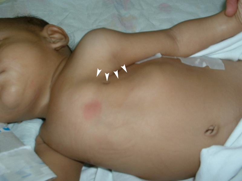
Patient's phenotype and marked thoracic depression (arrowheads).
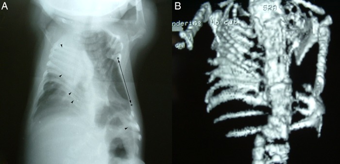
(A) Chest X-ray depicting ribs fanning out from their costovertebral joints (‘fan-like’ or ‘crab-like’ appearance) and a shortened spine with left lumbosacral scoliosis, altogether resulting in posterior rib fusions (arrowheads). A prominent intercostal hollow (*←→*) is seen, along with abnormal vertebral segmentation defects (‘pebble beach sign’) at the corresponding spinal segments. These findings characterise the spondylothoracic dysplasia subtype of JLS; notice the absence of intrinsic rib abnormalities (ie, aberrant rib count, bifurcations, thickenings, or more anteriorly located costal fusions). (B) Three-dimensional CT reconstruction of the same patient. JLS, Jarcho-Levin syndrome.
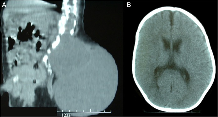
(A) Sagittal CT reconstruction defining contiguous left laminae defects and a large lumbosacral meningocele. Arachnoidal septations are seen within the meningocele's sac. (B) Head CT is normal in most cases of JLS, but should be performed to rule out other congenital syndromes. JLS, Jarcho-Levin syndrome.
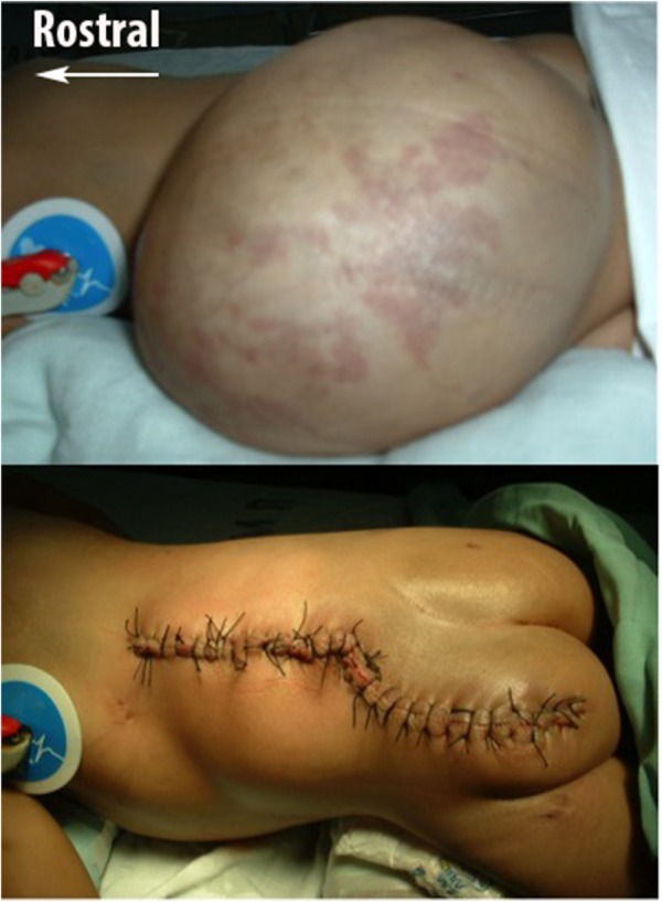
Upper: Preoperative photograph of the left-sided meningocele with fragile superficial vessels. Lower: Postoperative photograph of the final skin closure.
Similar articles
-
[Description of a case of spondylo-thoracic dysplasia or Jarcho-Levin syndrome].Pediatr Med Chir. 1998 Sep-Oct;20(5):353-5. Pediatr Med Chir. 1998. PMID: 10068987 Italian.
-
Controversies surrounding Jarcho-Levin syndrome.Curr Opin Pediatr. 2003 Dec;15(6):614-20. doi: 10.1097/00008480-200312000-00012. Curr Opin Pediatr. 2003. PMID: 14631208 Review.
-
Second trimester prenatal diagnosis of the Jarcho-Levin syndrome.Prenat Diagn. 1987 Feb;7(2):129-34. doi: 10.1002/pd.1970070209. Prenat Diagn. 1987. PMID: 3554211
-
Ten year follow-up of Jarcho-Levin syndrome with thoracic insufficiency treated by VEPTR and MCGR VEPTR hybrid.Eur Spine J. 2018 Jul;27(Suppl 3):287-291. doi: 10.1007/s00586-017-5164-x. Epub 2017 Jun 12. Eur Spine J. 2018. PMID: 28608177
-
Jarcho-levin syndrome associated with a complex congenital heart anomaly.Pediatr Cardiol. 2003 Jan-Feb;24(1):86-8. doi: 10.1007/s00246-002-1448-x. Pediatr Cardiol. 2003. PMID: 12574986 Review.
Cited by
-
Anatomical Engineering and 3D Printing for Surgery and Medical Devices: International Review and Future Exponential Innovations.Biomed Res Int. 2022 Mar 24;2022:6797745. doi: 10.1155/2022/6797745. eCollection 2022. Biomed Res Int. 2022. PMID: 35372574 Free PMC article. Review.
References
-
- Jarcho S, Levin PM. Hereditary malformation of the vertebral bodies. Bull John Hopkins Hosp 1938;62:216–26.
-
- Turnpenny PD, Young E. Icvas. Spondylocostal Dysostosis, Autosomal Recessive. In: Pagon RA, Adam MP, Ardinger HH et al., eds. GeneReviews(R). Seattle, WA: University of Washington, Seattle; All rights reserved, 1993.
Publication types
MeSH terms
Supplementary concepts
LinkOut - more resources
Full Text Sources
Other Literature Sources