. 2015 Sep;21(9):1648-59.
doi: 10.1261/rna.052464.115. Epub 2015 Jul 21.

O6-Methylguanosine leads to position-dependent effects on ribosome speed and fidelity

Affiliations

O6-Methylguanosine leads to position-dependent effects on ribosome speed and fidelity

Benjamin H Hudson et al. RNA. 2015 Sep.

Abstract

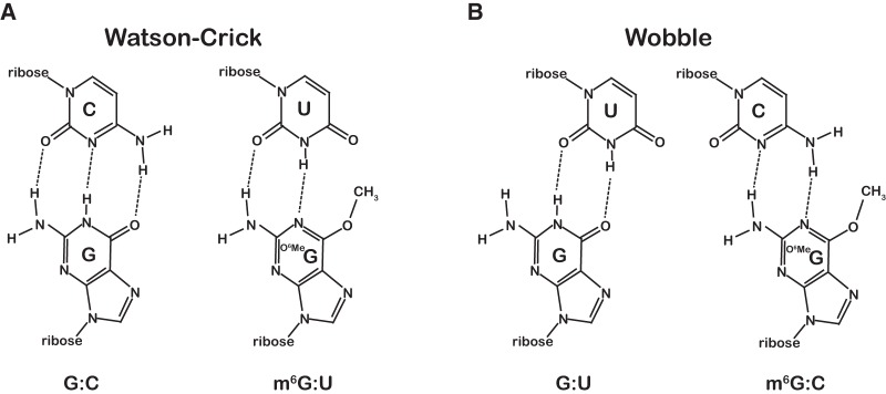
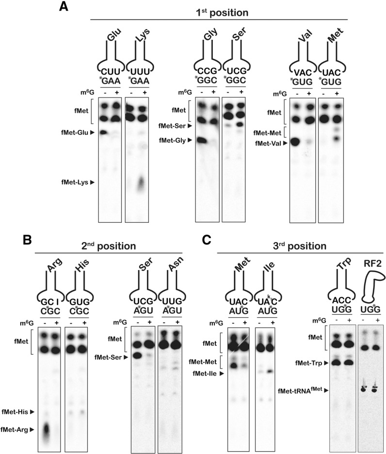
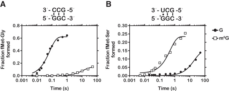
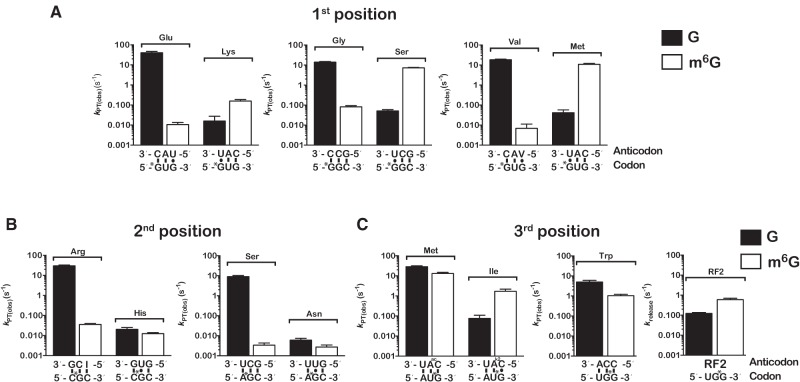
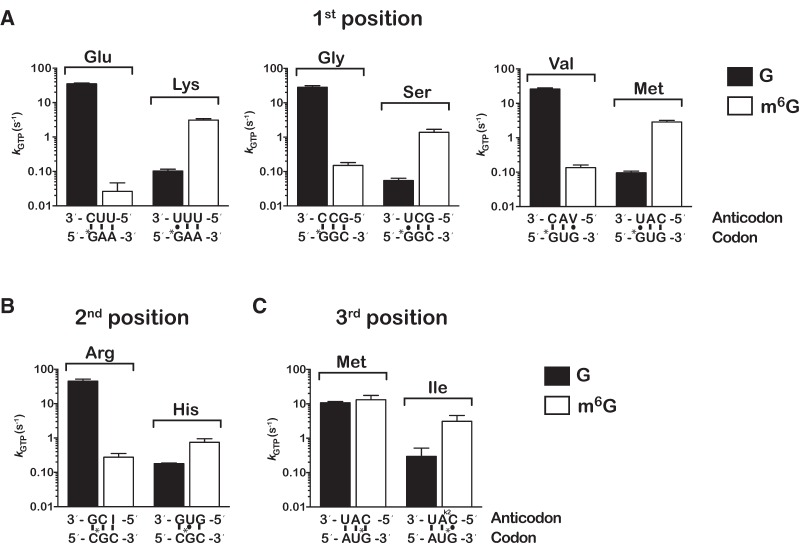
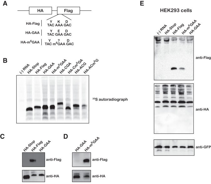
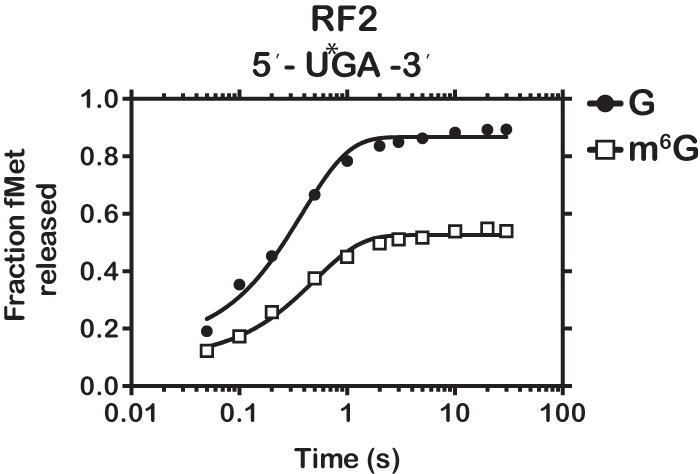
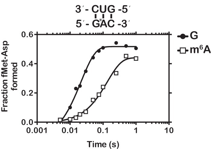
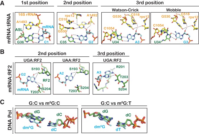
Nucleic acids are under constant assault from endogenous and environmental agents that alter their physical and chemical properties. O6-methylation of guanosine (m(6)G) is particularly notable for its high mutagenicity, pairing with T, during DNA replication. Yet, while m(6)G accumulates in both DNA and RNA, little is known about its effects on RNA. Here, we investigate the effects of m(6)G on the decoding process, using a reconstituted bacterial translation system. m(6)G at the first and third position of the codon decreases the accuracy of tRNA selection. The ribosome readily incorporates near-cognate aminoacyl-tRNAs (aa-tRNAs) by forming m(6)G-uridine codon-anticodon pairs. Surprisingly, the introduction of m(6)G to the second position of the codon does not promote miscoding, but instead slows the observed rates of peptide-bond formation by >1000-fold for cognate aa-tRNAs without altering the rates for near-cognate aa-tRNAs. These in vitro observations were recapitulated in eukaryotic extracts and HEK293 cells. Interestingly, the analogous modification N6-methyladenosine (m(6)A) at the second position has only a minimal effect on tRNA selection, suggesting that the effects on tRNA selection seen with m(6)G are due to altered geometry of the base pair. Given that the m6G:U base pair is predicted to be nearly indistinguishable from a Watson-Crick base pair, our data suggest that the decoding center of the ribosome is extremely sensitive to changes at the second position. Our data, apart from highlighting the deleterious effects that these adducts pose to cellular fitness, shed new insight into decoding and the process by which the ribosome recognizes codon-anticodon pairs.

Keywords: O6-methylguanosine; RNA damage; decoding; ribosome; translation.


Figures

FIGURE 1.
FIGURE 1.
m6G:U base pair adopts a conformation similar to a Watson-Crick base pair. (A) Chemical structure of a normal Watson-Crick G:C base pair compared to the mutagenic m6G:U base pair structure. (B) Chemical structures of the G:U and m6G:C wobble base pairs.
FIGURE 2.
FIGURE 2.
m6G affects the accuracy and speed of the ribosome. (AC) Phosphorimager scans of electrophoretic TLCs used to follow dipeptide formation in the presence of the indicated initiation and ternary complexes.
FIGURE 3.
FIGURE 3.
Representative time courses of cognate and near-cognate dipeptide formation. (A) Time course of peptide-bond formation between the native GGC (closed circle), methylated m6GGC (closed square), and cognate Gly-tRNAGly ternary complex. (B) Time course of peptide-bond formation between the native GGC (closed circle), methylated m6GGC (closed square), and near-cognate Ser-tRNASer ternary complex.
FIGURE 4.
FIGURE 4.
m6G affects the decoding process in a position-dependent manner. (A) Bar graphs of the observed rates of peptide-bond formation for complexes carrying m6G at the first position of the codon relative to their native counterparts. For each pair of complexes, rates measured in the presence of cognate ternary complexes are plotted in the left two bars in each graph, whereas those measured in the presence of the near-cognate ternary complexes (U:G at the first position) are plotted in the right two bars. The codon–anticodon interactions are depicted below the x-axis with the corresponding dipeptide depicted above the bars. (B) Same as A, but with initiation complexes harboring m6G at the second position of the A-site codon. (C) Same as A, but with initiation complexes harboring m6G at the third position of the A-site codon. Note that the graph for the UGG and UGm6G complexes was split into two. Error bars represent the standard error of curve fitting from a single representative time course. Twenty-three of the 28 time courses were performed in duplicates with at least <10% variability between samples.
FIGURE 5.
FIGURE 5.
m6G alters the observed rate of GTP hydrolysis by EF-Tu. (A) Observed rates of GTP hydrolysis measured on native and the corresponding first-position modified complexes with cognate (left two bars in each graph) and near-cognate ternary complexes (right two bars in each graph). Labeling scheme of the graphs is identical to the one used in Figure 4. (B,C) Similar to A, but with complexes programmed with m6G at the second position and third position of the codon, respectively. Error bars represent the standard error of curve fit from a single representative time course.
FIGURE 6.
FIGURE 6.
Effects of m6G on translation in eukaryotic extracts and mammalian cell culture. (A) Schematic of model reporter mRNAs depicting N-terminal HA tag, C-terminal Flag tag, and single m6G residue. (B) Phosphorimage scan of 35S-labeled peptide products after wheat germ extract translation. (C) Immunoblots of wheat germ translated peptides probed with anti-Flag and anti-HA antibodies. (D) Same as in C, but comparing the HA-GAA and HA-m6GAA mRNAs. (E) Immunoblots of HEK293T lysates transfected with m6G reporter mRNAs along with control GFP plasmid and probed with anti-Flag, anti-HA, and anti-GFP antibodies.
FIGURE 7.
FIGURE 7.
m6G has little effect on peptide release. Time course of RF2-dependent fMet release for initiation complexes programmed with UGA or Um6GA.
FIGURE 8.
FIGURE 8.
m6A has a marginal effect on the rate of peptide-bond formation. Time course of peptide-bond formation between initiation complexes programmed with GAC or the m6G-related modification Gm6AC and the cognate Asp-tRNAAsp ternary complex.
FIGURE 9.
FIGURE 9.
Recognition of base-pairing geometry by the ribosome and a high-fidelity DNA polymerase. (A) Inspection of the minor groove of the codon–anticodon pair by 16S rRNA and rps12 of the small subunit. (PDB IDs: 1XNR and 2WDG) (Murphy and Ramakrishnan 2004; Voorhees et al. 2009). (B) Recognition of stop codons by RF2 at the second position and third position (PDB IDs: 2WH3 and 4V67) (Korostelev et al. 2008; Laurberg et al. 2008; Weixlbaumer et al. 2008). (C) Structural alignments of a dm6G:dC base pair (PDB ID: 2HVH, left) or a dm6G:dT base pair (PDB ID: 2HVW, right) onto a dG:dC base pair (PDB ID: 2HVI) in the active site of a high-fidelity DNA polymerase (Warren et al. 2006). All molecular representations were generated using PyMol (DeLano Scientific).

References

    1. Aas PA, Otterlei M, Falnes PO, Vågbø CB, Skorpen F, Akbari M, Sundheim O, Bjørås M, Slupphaug G, Seeberg E, et al. 2003. Human and bacterial oxidative demethylases repair alkylation damage in both RNA and DNA. Nature 421: 859–863. - PubMed
    1. Carter AP, Clemons WM Jr, Brodersen DE, Morgan-Warren RJ, Hartsch T, Wimberly BT, Ramakrishnan V. 2001. Crystal structure of an initiation factor bound to the 30S ribosomal subunit. Science 291: 498–501. - PubMed
    1. Demeshkina N, Jenner L, Westhof E, Yusupov M, Yusupova G. 2012. A new understanding of the decoding principle on the ribosome. Nature 484: 256–259. - PubMed
    1. Demple B, Jacobsson A, Olsson M, Robins P, Lindahl T. 1982. Repair of alkylated DNA in Escherichia coli. Physical properties of O6-methylguanine-DNA methyltransferase. J Biol Chem 257: 13776–13780. - PubMed
    1. Doma MK, Parker R. 2006. Endonucleolytic cleavage of eukaryotic mRNAs with stalls in translation elongation. Nature 440: 561–564. - PMC - PubMed

Publication types

LinkOut - more resources