

Acrylamide neurotoxicity on the cerebrum of weaning rats
- PMID: 26199611
- PMCID: PMC4498356
- DOI: 10.4103/1673-5374.158357
Acrylamide neurotoxicity on the cerebrum of weaning rats
Abstract
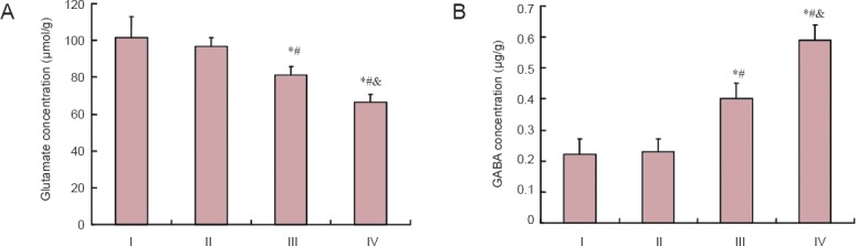
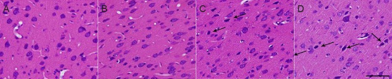
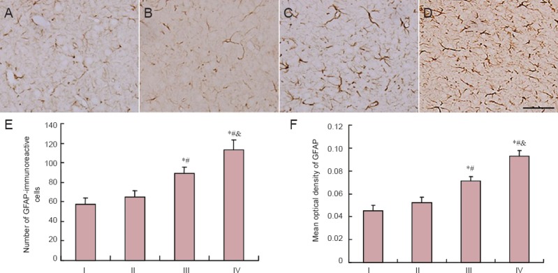
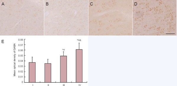
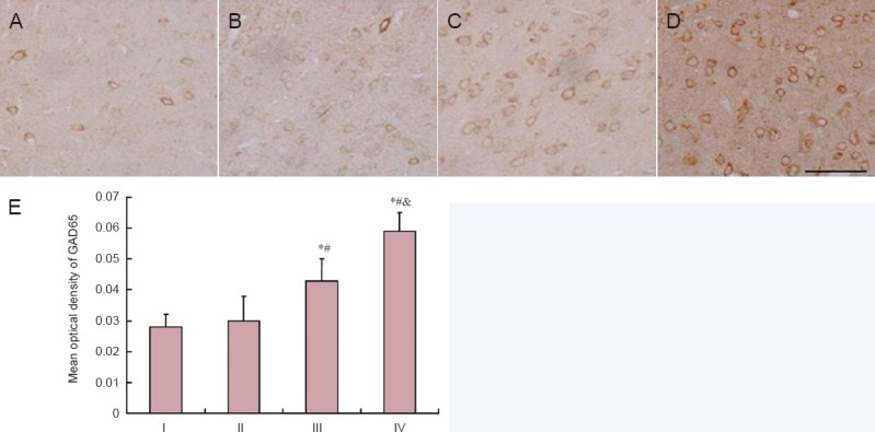
The mechanism underlying acrylamide-induced neurotoxicity remains controversial. Previous studies have focused on acrylamide-induced toxicity in adult rodents, but neurotoxicity in weaning rats has not been investigated. To explore the neurotoxic effect of acrylamide on the developing brain, weaning rats were gavaged with 0, 5, 15, and 30 mg/kg acrylamide for 4 consecutive weeks. No obvious neurotoxicity was observed in weaning rats in the low-dose acrylamide group (5 mg/kg). However, rats from the moderate- and high-dose acrylamide groups (15 and 30 mg/kg) had an abnormal gait. Furthermore, biochemical tests in these rats demonstrated that glutamate concentration was significantly reduced, and γ-aminobutyric acid content was significantly increased and was dependent on acrylamide dose. Immunohistochemical staining showed that in the cerebral cortex, γ-aminobutyric acid, glutamic acid decarboxylase and glial fibrillary acidic protein expression increased remarkably in the moderate- and high-dose acrylamide groups. These results indicate that in weaning rats, acrylamide is positively associated with neurotoxicity in a dose-dependent manner, which may correlate with upregulation of γ-aminobutyric acid and subsequent neuronal degeneration after the initial acrylamide exposure.
Keywords: cerebrum; cortex; glial fibrillary acidic protein; glutamate; glutamic acid decarboxylase; nerve regeneration; neural regeneration; neurotoxicity; organ index; weaning; γ-aminobutyric acid.
Conflict of interest statement
Figures
Similar articles
-
Toxic effect of acrylamide on the development of hippocampal neurons of weaning rats.Neural Regen Res. 2017 Oct;12(10):1648-1654. doi: 10.4103/1673-5374.217345. Neural Regen Res. 2017. PMID: 29171430 Free PMC article.
-
Effect of sub-acute exposure to acrylamide on GABAergic neurons and astrocytes in weaning rat cerebellum.Toxicol Ind Health. 2012 Feb;28(1):10-20. doi: 10.1177/0748233711401264. Epub 2011 Mar 28. Toxicol Ind Health. 2012. PMID: 21444355
-
The Possible Protective Role of Dark Chocolate Against Acrylamide Neurotoxicity in Weaning Rats Cerebellum.Mol Neurobiol. 2022 Jan;59(1):234-244. doi: 10.1007/s12035-021-02580-x. Epub 2021 Oct 18. Mol Neurobiol. 2022. PMID: 34661852
-
Amended final report on the safety assessment of polyacrylamide and acrylamide residues in cosmetics.Int J Toxicol. 2005;24 Suppl 2:21-50. doi: 10.1080/10915810590953842. Int J Toxicol. 2005. PMID: 16154914 Review.
-
Acrylamide neurotoxicity.Nutr Neurosci. 2014 Feb;17(2):49-57. doi: 10.1179/1476830513Y.0000000065. Epub 2013 Nov 26. Nutr Neurosci. 2014. PMID: 23541332 Review.
Cited by
-
Developmental and Neurotoxicity of Acrylamide to Zebrafish.Int J Mol Sci. 2021 Mar 29;22(7):3518. doi: 10.3390/ijms22073518. Int J Mol Sci. 2021. PMID: 33805345 Free PMC article.
-
Effects of acrylamide exposure during pregnancy and lactation on the development of myelin sheath of corpus callosum in offspring rats.Toxicol Res (Camb). 2024 Feb 1;13(1):tfae014. doi: 10.1093/toxres/tfae014. eCollection 2024 Feb. Toxicol Res (Camb). 2024. PMID: 38314039 Free PMC article.
-
AQP4 Attenuated TRAF6/NFκB Activation in Acrylamide-Induced Neurotoxicity.Molecules. 2022 Feb 4;27(3):1066. doi: 10.3390/molecules27031066. Molecules. 2022. PMID: 35164330 Free PMC article.
-
Acrylamide acute neurotoxicity in adult zebrafish.Sci Rep. 2018 May 21;8(1):7918. doi: 10.1038/s41598-018-26343-2. Sci Rep. 2018. PMID: 29784925 Free PMC article.
-
Toxic effect of acrylamide on the development of hippocampal neurons of weaning rats.Neural Regen Res. 2017 Oct;12(10):1648-1654. doi: 10.4103/1673-5374.217345. Neural Regen Res. 2017. PMID: 29171430 Free PMC article.
References
-
- Agrawal AK, Seth PK, Squibb RE, Tilson HA, Uphouse LL, Bondy SC. Neurotransmitter receptors in brain regions of acrylamide-treated rats. I: Effects of a single exposure to acrylamide. Pharmacol Biochem Behav. 1981;14:527–531. - PubMed
-
- Bao X, Chu S. Beijing: People's Medical Publishing House; 1991. The Rat Barin Atlas.
-
- Bondy SC, Tilson HA, Agrawal AK. Neurotransmitter receptors in brain regions of acrylamide-treated rats. II: effects of extended exposure to acrylamide. Pharmacol Biochem Behav. 1981;14:533–537. - PubMed
-
- Costa LG, Deng H, Gregotti C, Manzo L, Faustman EM, Bergmark E, Calleman CJ. Comparative studies on the neuro and reproductive toxicity of acrylamide and its epoxide metabolite glycidamide in the rat. Neurotoxicology. 1992;13:219–224. - PubMed
-
- Deng L, Yuan Q. The study progress of GFAP in the neuronal system. Luzhou Yixueyuan Xuebao. 2005;2:189–192.
LinkOut - more resources
Full Text Sources
Other Literature Sources