Review
. 2015:2015:517520.
doi: 10.1155/2015/517520. Epub 2015 Jun 25.

Advances and Prospects in Tissue-Engineered Meniscal Scaffolds for Meniscus Regeneration

Affiliations
Review

Advances and Prospects in Tissue-Engineered Meniscal Scaffolds for Meniscus Regeneration

Weimin Guo et al. Stem Cells Int. 2015.

Abstract

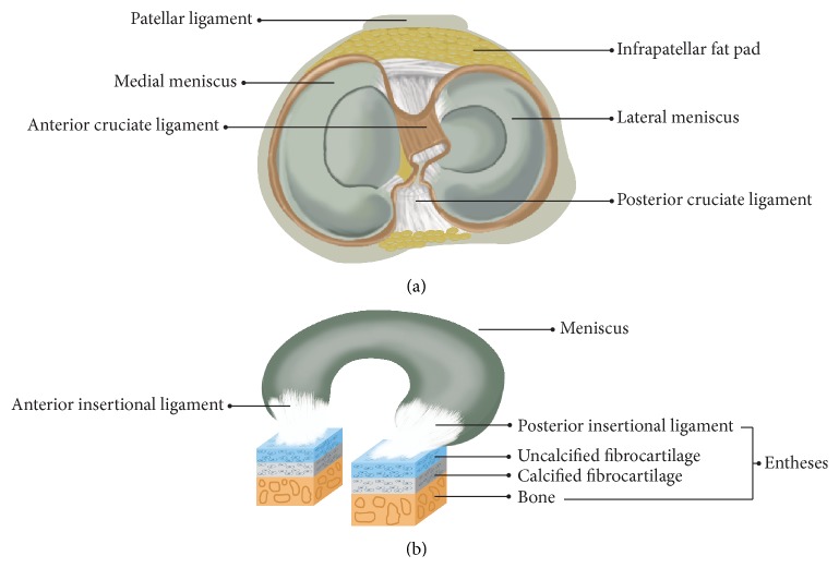
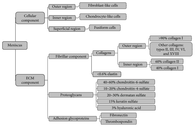
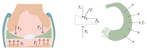
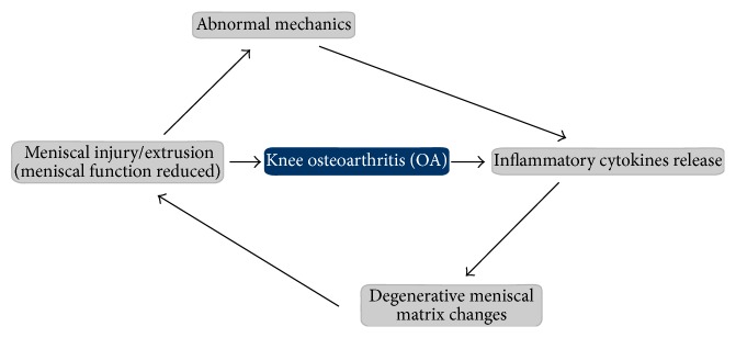
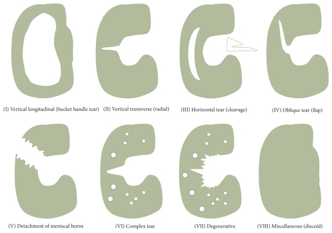
The meniscus plays a crucial role in maintaining knee joint homoeostasis. Meniscal lesions are relatively common in the knee joint and are typically categorized into various types. However, it is difficult for inner avascular meniscal lesions to self-heal. Untreated meniscal lesions lead to meniscal extrusions in the long-term and gradually trigger the development of knee osteoarthritis (OA). The relationship between meniscal lesions and knee OA is complex. Partial meniscectomy, which is the primary method to treat a meniscal injury, only relieves short-term pain; however, it does not prevent the development of knee OA. Similarly, other current therapeutic strategies have intrinsic limitations in clinical practice. Tissue engineering technology will probably address this challenge by reconstructing a meniscus possessing an integrated configuration with competent biomechanical capacity. This review describes normal structure and biomechanical characteristics of the meniscus, discusses the relationship between meniscal lesions and knee OA, and summarizes the classifications and corresponding treatment strategies for meniscal lesions to understand meniscal regeneration from physiological and pathological perspectives. Last, we present current advances in meniscal scaffolds and provide a number of prospects that will potentially benefit the development of meniscal regeneration methods.


Figures

Figure 1
Figure 1
Top view of the anatomical meniscus (a): lateral meniscus is “O” shaped, whereas the medial meniscus has a “C” appearance. The meniscal functional unit (b), including the corresponding anterior and posterior ligaments and entheses. Entheses typically contain ligaments, uncalcified fibrocartilage, calcified fibrocartilage, and bone.
Figure 2
Figure 2
The complex composition of the meniscal cellular and meniscal extracellular matrix (ECM) components. The outer region is the outer one-third of the meniscus; the inner region is the inner two-thirds of the meniscus; the superficial region is the surface of the meniscus.
Figure 3
Figure 3
Schematic diagram of the meniscus force-bearing mechanism. Meniscal configuration adapts well to the corresponding shape of the femoral condyles and the tibial plateau in the knee joint. The axial load force (F) perpendicular to the meniscus surface and horizontal force (f r) are created by compressing the femur (F f). F rebounds due to the tibial upgrade force (F t), whereas the f r leads to meniscal extrusion radially, which is countered by the pulling force from the anterior and posterior insertional ligaments. Consequently, tensile hoop stress is created along the circumferential directions during axial compression.
Figure 4
Figure 4
The interaction between meniscal injury and knee osteoarthritis (OA). Knee mechanics become abnormal when a meniscus is injured, which leads to increasing stress across adjacent cartilage and subchondral bone. This stress triggers release of inflammatory cytokines, which further impair the meniscal extracellular matrix (ECM) and accelerate the vicious cycle of knee OA. OA of the knee joint also causes release of inflammatory cytokines, repeating the vicious cycle.
Figure 5
Figure 5
Schematic diagram of the eight different types of meniscal lesions according to Casscells classification.

References

    1. Scotti C., Hirschmann M. T., Antinolfi P., Martin I., Peretti G. M. Meniscus repair and regeneration: review on current methods and research potential. European Cells & Materials. 2013;26:150–170. - PubMed
    1. Baker B. E., Peckham A. C., Pupparo F., Sanborn J. C. Review of meniscal injury and associated sports. The American Journal of Sports Medicine. 1985;13(1):1–4. doi: 10.1177/036354658501300101. - DOI - PubMed
    1. Hede A., Jensen D. B., Blyme P., Sonne-Holm S. Epidemiology of meniscal lesions in the knee: 1,215 open operations in Copenhagen 1982–84. Acta Orthopaedica. 1990;61(5):435–437. doi: 10.3109/17453679008993557. - DOI - PubMed
    1. Annandale T. Excision of the internal semilunar cartilage, resulting in perfect restoration of the joint-movements. The British Medical Journal. 1889;1(1467):291–292. doi: 10.1136/bmj.1.1467.291. - DOI - PMC - PubMed
    1. Gear M. W. The late results of meniscectomy. The British Journal of Surgery. 1967;54(4):270–272. doi: 10.1002/bjs.1800540406. - DOI - PubMed