. 2015 Jul 21:3:32.
doi: 10.1186/s40425-015-0077-x. eCollection 2015.

Route of antigen delivery impacts the immunostimulatory activity of dendritic cell-based vaccines for hepatocellular carcinoma

Affiliations

Route of antigen delivery impacts the immunostimulatory activity of dendritic cell-based vaccines for hepatocellular carcinoma

Angela D Pardee et al. J Immunother Cancer. .

Abstract

Background: Dendritic cells (DC) are uniquely equipped to capture, process, and present antigens from their environment. The context in which an antigen is acquired by DC helps to dictate the subsequent immune response. Cancer vaccination promotes antitumor immunity by directing an immune response to antigens expressed by tumors. We have tested the tumor-associated antigen alpha-fetoprotein (AFP) as an immunotherapy target. The majority of hepatocellular carcinomas (HCC) upregulate and secrete this oncofetal antigen.

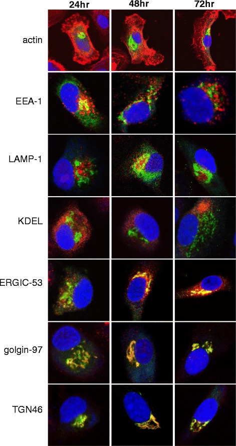
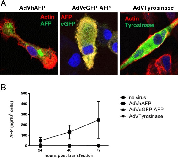
Methods: To develop cancer vaccines for HCC capable of promoting potent tumor-specific T cell responses, we tested adenovirally-encoded synthetic AFP, with or without its signal sequence, as well as protein forms of AFP and compared intracellular routing and subsequent antigen-specific CD8+ and CD4+ T cell responses.

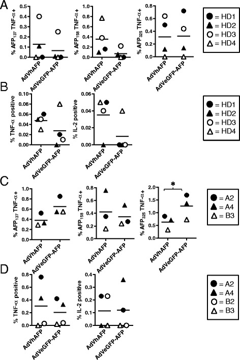
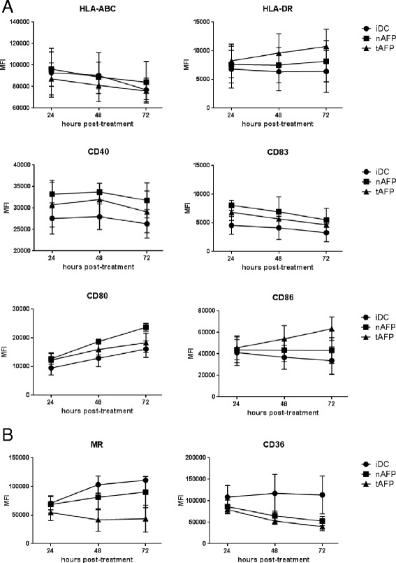
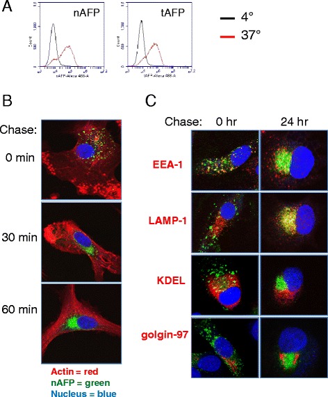
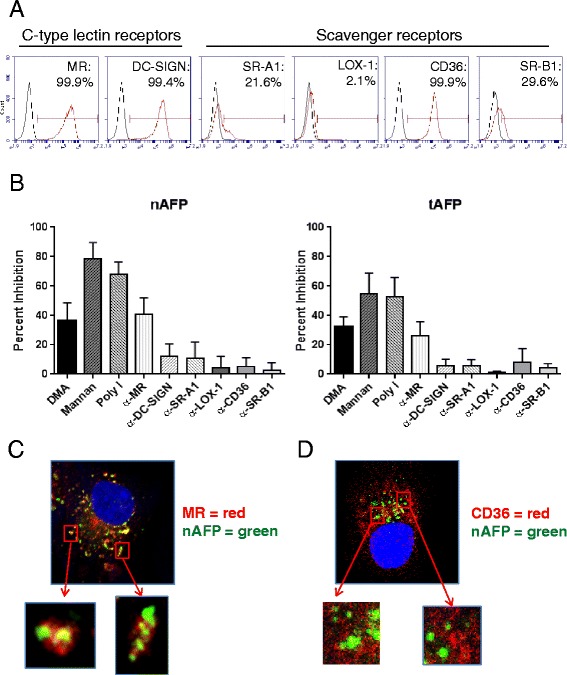
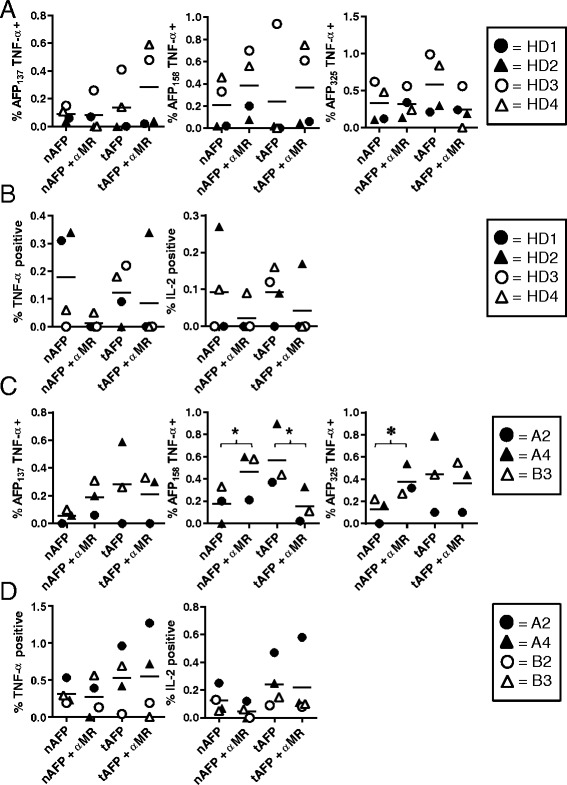
Results: Surprisingly, the secreted form of antigen was superior for both CD4+ and CD8+ T cell activation. We also examined the mechanism through which AFP protein is endocytosed and trafficked in human DC. We identify the mannose receptor (MR/CD206) as the primary uptake pathway for both normal cord blood-derived AFP (nAFP) and tumor-derived AFP (tAFP) proteins. While in healthy donors, nAFP and tAFP were cross-presented to CD8+ T cells similarly and CD4+ T cell responses were dependent upon MR-mediated uptake. In HCC patient cells, tAFP was more immunogenic, and CD4+ T cell responses were not MR-dependent.

Conclusions: Secreted, cytoplasmically retained, and endocytosed forms of AFP utilize unique uptake and processing pathways, resulting in different immunologic responses from the induced antigen-specific CD4+ and CD8+ T cells and between healthy donors and HCC patients. Collectively, these data elucidate pathways of spontaneous and induced anti-tumor immunity in HCC patients to this secreted antigen.

Keywords: Adenovirus; Alpha-fetoprotein; Dendritic cells; Hepatocellular carcinoma.


Figures

Fig. 1
Fig. 1
Phenotype of AdV-transduced DC. a and (b) Immature DC (iDC) from healthy donors (n = 3) were left untreated, matured with LPS/IFN-γ (mDC), or transduced with AdVhAFP, and then cultured in DC media for 24, 48, or 72 hr. Cells were stained for (a) antigen presentation and costimulatory markers and (b) endocytic receptors, and analyzed by flow cytometry. Mean fluorescence intensity (MFI) is reported as the mean ± SD
Fig. 2
Fig. 2
Intracellular trafficking of adenovirally-expressed AFP. DC were transduced with AdVhAFP at MOI 2000 for 3 hr, and then cultured in DC media for 24, 48, or 72 hr. Cells were then fixed and stained for AFP (green), actin, EEA-1 (early endosomes), LAMP-1 (late endosomes/lysosomes), KDEL (endoplasmic reticulum/ER), ERGIC-53 (ER-Golgi intermediate complex), golgin-97 (Golgi), and TGN46 (trans-Golgi network) (all in red), as described in Materials and Methods. All images are representative of three independent experiments performed and were taken using a 63x objective
Fig. 3
Fig. 3
Adenovirally-expressed AFP, but not eGFP-AFP, is secreted by transduced DC. a DC were transduced with AdVhAFP (left panel), AdVeGFP-AFP (middle panel), or AdVTyrosinase at MOI 2000 for 3 hr. After 48 hr, cells were fixed and stained for AFP (left and middle panels), actin (left and right panels), or Tyrosinase (right panel). Antibody staining of AdVeGFP-AFP-transduced cells with an anti-AFP antibody reveals that AFP and GFP colocalize, as expected. Representative images from three independent experiments are shown. Images were taken using a 63x objective. b DC from healthy donors (n = 3) were transduced as above and cultured in DC media for 24, 48, or 72 hr. Supernatants were analyzed by AFP ELISA. Data are reported as the mean ± SD
Fig. 4
Fig. 4
Ability of AdV-transduced DC to induce AFP-specific T cell responses. a and (b) DC from HLA-A2+ HDs (n = 4) were transduced with AdVhAFP or AdVeGFP-AFP at MOI 2000 for 3 hr, then co-cultured with autologous PBMC for 12–13 days. a CD8+ T cells were restimulated with autologous DC (loaded with AFP as in the initial stimulation) for an additional 10d, and then analyzed for AFP-specific intracellular TNF-α production against three immunodominant HLA-A2-restricted peptides. b CD4+ T cells were collected after the initial stimulation and analyzed for AFP-specific intracellular cytokine production. (C and D) DC from HLA-A2+ HCC patients (n = 3–4) were transduced with AdVhAFP or AdVeGFP-AFP at MOI 2000 for 3 hr, then co-cultured with autologous PBMC for 12–13 days. c CD8+ T cells were collected and analyzed for AFP-specific intracellular TNF-α production against three immunodominant HLA-A2-restricted peptides. d CD4+ T cells were collected and analyzed for AFP-specific intracellular cytokine production. For each panel, the solid lines represent the mean value. *, P < 0.05
Fig. 5
Fig. 5
Phenotype of AFP-loaded DC. a and (b) Immature DC (iDC) from healthy donors (n = 3) were left untreated or cultured with nAFP and tAFP (10 μg/ml) in DC media for 24, 48, or 72 hr. Cells were stained for (a) antigen presentation and costimulatory markers and (b) endocytic receptors, and analyzed by flow cytometry. Mean fluorescence intensity (MFI) is reported as the mean ± SD
Fig. 6
Fig. 6
Intracellular trafficking of endocytosed AFP. a nAFP and tAFP were Alexa Fluor 488-labeled. Human monocyte-derived DC were co-cultured with AFP (10 μg/ml) for 1 hr at 4 °C or 37 °C, and analyzed by flow cytometry. b DC were co-cultured with fluorescently-labeled nAFP for 2 hr, then chased with media alone for the indicated times. Cells were then fixed, stained for actin (described in Materials and Methods), and analyzed by confocal microscopy. c DC were incubated with fluorescently-labeled nAFP for 2 hr, then chased with media alone for the indicated times. Cells were then fixed and stained for EEA-1 (early endosomes), LAMP-1 (late endosomes/lysosomes), KDEL (endoplasmic reticulum/ER), and golgin-97 (Golgi) (all in red). All images are representative of three independent experiments performed and were taken using a 63x objective
Fig. 7
Fig. 7
DC endocytose AFP primarily via the mannose receptor. a DC were stained for flow cytometric analysis. Red histogram represents endocytic receptor staining; black histogram represents isotype control staining. Data from one representative healthy donor (of three total HD tested) is shown. b DC were pre-treated with inhibitors for 30 min, and co-cultured with fluorescently-labeled nAFP (top panels) or tAFP (bottom panels) for an additional 1 hr. Percent inhibition was calculated based on untreated control cells. Columns, mean of four HD; bars, standard deviation. c and (d) DC were incubated with fluorescently-labeled nAFP for 2 hr, then fixed and stained for MR (c) or CD36 (d). All images are representative of three independent experiments performed and were taken using a 63x objective
Fig. 8
Fig. 8
Ability of AFP protein-loaded DC to induce AFP-specific T cell responses. a and (b) DC from HLA-A2+ HDs (n = 4) were cultured with nAFP or tAFP (at 10 μg/ml for 2 hr) with or without pre-treatment with MR blocking antibody (at 10 μg/ml for 30 min), matured for an additional 24 h, then co-cultured with autologous PBMC for 12–13 days. a CD8+ T cells were restimulated with autologous DC (loaded with AFP as in the initial stimulation) for an additional 10d, and then analyzed for AFP-specific intracellular TNF-α production against three immunodominant HLA-A2-restricted peptides. b CD4+ T cells were collected after the initial stimulation and analyzed for AFP-specific intracellular cytokine production. c and (d) DC from HLA-A2+ HCC patients (n = 3–4) were cultured with nAFP or tAFP (at 10 μg/ml for 2 hr) with or without pre-treatment with MR blocking antibody (at 10 μg/ml for 30 min), matured for an additional 24 h, then co-cultured with autologous PBMC for 12–13 days. (C) CD8+ T cells were collected and analyzed for AFP-specific intracellular TNF-α production against three immunodominant HLA-A2-restricted peptides. (D) CD4+ T cells were collected and analyzed for AFP-specific intracellular cytokine production. For each panel, the solid lines represent the mean value. *, P < 0.05

References

    1. Blander JM, Medzhitov R. Regulation of phagosome maturation by signals from toll-like receptors. Science. 2004;304(5673):1014–8. doi: 10.1126/science.1096158. - DOI - PubMed
    1. Burgdorf S, Kurts C. Endocytosis mechanisms and the cell biology of antigen presentation. Curr Opin Immunol. 2008;20(1):89–95. doi: 10.1016/j.coi.2007.12.002. - DOI - PubMed
    1. Sallusto F, Cella M, Danieli C, Lanzavecchia A. Dendritic cells use macropinocytosis and the mannose receptor to concentrate macromolecules in the major histocompatibility complex class II compartment: downregulation by cytokines and bacterial products. J Exp Med. 1995;182(2):389–400. doi: 10.1084/jem.182.2.389. - DOI - PMC - PubMed
    1. Fehres CM, Unger WW, Garcia-Vallejo JJ, van Kooyk Y. Understanding the biology of antigen cross-presentation for the design of vaccines against cancer. Front Immunol. 2014;5:149. doi: 10.3389/fimmu.2014.00149. - DOI - PMC - PubMed
    1. Burgdorf S, Kautz A, Bohnert V, Knolle PA, Kurts C. Distinct pathways of antigen uptake and intracellular routing in CD4 and CD8 T cell activation. Science. 2007;316(5824):612–6. doi: 10.1126/science.1137971. - DOI - PubMed

LinkOut - more resources