Review
. 2015 Jun 30:5:38.
doi: 10.4103/2156-7514.159565. eCollection 2015.

The Dens: Normal Development, Developmental Variants and Anomalies, and Traumatic Injuries

Affiliations
Review

The Dens: Normal Development, Developmental Variants and Anomalies, and Traumatic Injuries

William T O'Brien Sr et al. J Clin Imaging Sci. .

Abstract

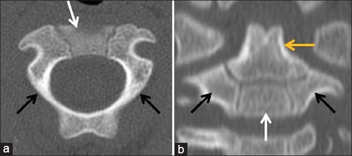
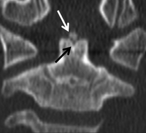
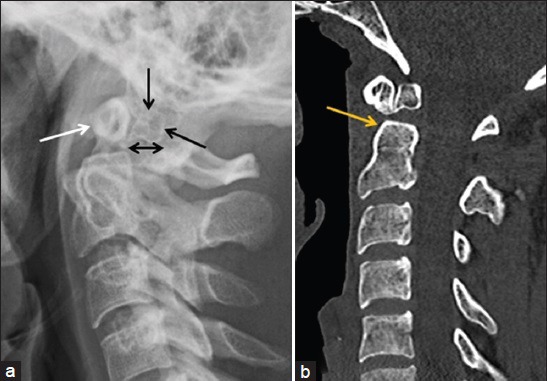
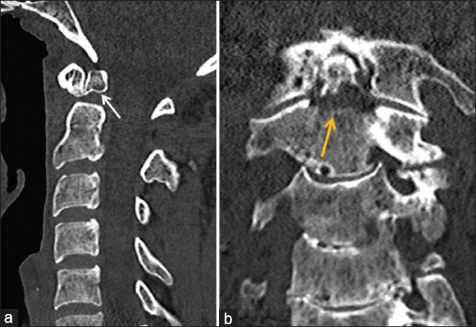
Accurate interpretation of cervical spine imagining can be challenging, especially in children and the elderly. The biomechanics of the developing pediatric spine and age-related degenerative changes predispose these patient populations to injuries centered at the craniocervical junction. In addition, congenital anomalies are common in this region, especially those associated with the axis/dens, due to its complexity in terms of development compared to other vertebral levels. The most common congenital variations of the dens include the os odontoideum and a persistent ossiculum terminale. At times, it is necessary to distinguish normal development, developmental variants, and developmental anomalies from traumatic injuries in the setting of acute traumatic injury. Key imaging features are useful to differentiate between traumatic fractures and normal or variant anatomy acutely; however, the radiologist must first have a basic understanding of the spectrum of normal developmental anatomy and its anatomic variations in order to make an accurate assessment. This review article attempts to provide the basic framework required for accurate interpretation of cervical spine imaging with a focus on the dens, specifically covering the normal development and ossification of the dens, common congenital variants and their various imaging appearances, fracture classifications, imaging appearances, and treatment options.

Keywords: Cervical vertebrae; dens axis; odontoid process; spinal anatomy; spinal injuries.


Figures

Figure 1
Figure 1
38-month-old child with normal ossification centers. (a) Axial CT image through the base of the dens shows the ossification centers of the lateral neural arches (black arrows) and anterior body (white arrow) of the axis. The posterior neural arches have already fused. (b) Coronal reformatted CT image through the anterior aspect of the C-2 vertebral body shows the lateral neural arch. (black arrows) and anterior body. (white) ossification centers, as well as the ossification center of the dens superiorly. (yellow arrow). The ossiculum terminale has yet to ossify.
Figure 2
Figure 2
7-year-old child with a normal ossiculum terminale. Coronal reformatted CT image through the anterior aspect of the C-2 vertebral body shows ossification of the ossiculum terminale along the superior margin of the dens (white arrow). A characteristic “V”-shaped cartilaginous cleft is seen underlying the ossiculum terminale (black arrow).
Figure 3
Figure 3
26-year-old man with chronic neck pain and an os odontoideum. (a) Lateral radiograph of the cervical spine shows posterior displacement of a well-corticated os odontoideum (black arrows), as well as hypertrophy of the anterior arch of C-1 (white arrow). (b) Reformatted sagittal CT image in the same patient demonstrates similar findings with restoration of normal alignment in the supine position. The margins of the os odontoideum are smooth and well-corticated, and the base of dens is relatively foreshortened (yellow arrow). The variation in alignment with positioning and posterior displacement of the os odontoideum on the plain radiograph confirms instability.
Figure 4
Figure 4
26-year-old man with chronic neck pain and an os odontoideum versus an elderly woman with an acute Type. II dens fracture. (a) Reformatted sagittal CT image in the same young adult as Figure 3a demonstrates well-corticated margins of an os odontoideum. (white arrow) with characteristic secondary features, to include a hypertrophied anterior arch of C-1 and a relatively foreshortened base of the dens. (b) Coronal reformatted CT image through the upper cervical spine in an elderly woman with a fall demonstrates a fracture through the base of the dens with lucent margins. (yellow arrow).
Figure 5
Figure 5
28-year-old man with chronic neck pain and an os odontoideum with instability. (a) Sagittal T2 MR image through the cervical spine in the neutral position shows normal alignment of the os odontoideum with respect to the C-2 body and a normal atlanto-dental interval (yellow arrow). (b) Repeat imaging with passive flexion shows posterior displacement of the os odontoideum (white arrow) and widening of the atlanto-dental interval (yellow arrow).
Figure 6
Figure 6
10-year-old boy with a normal ossiculum terminale versus an elderly gentleman post-trauma with an acute Type I dens fracture. (a) Coned-down reformatted coronal CT image of the C-2 vertebral body in the young boy shows a smooth, corticated ossification along the superior margin of the dens (white arrow) with an underlying “V”-shaped cartilaginous cleft. (b) Sagittal reformatted CT image in the elderly trauma patient demonstrates an ossific fragment along the superior margin of the dens (white arrow) with a subjacent lucent fracture line (black arrow).
Figure 7
Figure 7
Illustration in the coronal plane demonstrates the odontoid fracture classification, as introduced by Anderson and D’Alonzo. Courtesy of Gilbert Gardner, Medical Illustrator.
Figure 8
Figure 8
Elderly gentleman post-trauma with neck pain and a Type I dens fracture. (a) Sagittal and. (b) coronal reformatted CT images through the cervical spine show an ossific fragment along the superior aspect of the dens (white arrows) with a subjacent lucent fracture line. (black arrows).
Figure 9
Figure 9
Middle-aged man post-trauma with multiple injuries, including a Type II dens fracture. (a) Sagittal reformatted CT image through the cervical spine demonstrates a displaced fracture through the base of the dens. (arrow) with small ossific fragments posteriorly. There is posterior displacement of the superior dens fragment and C-1 with respect to the base of the C-2 vertebral body. (b) Sagittal T2 MR image through the cervical spine shows the fracture. (arrow) with associated edema. (c) Lateral plain radiograph post internal anterior reduction and fixation demonstrates improved anatomic alignment.
Figure 10
Figure 10
Young child post-trauma with a dens fracture through the synchondrosis. (a) Lateral radiograph of the cervical spine demonstrates abnormal anterior displacement and angulation of the dens with respect to the body of C-2 (arrow). (b) Sagittal reformatted CT image shows similar findings with the injury occurring through the synchondrosis (arrow). (c) Lateral radiograph post non-surgical external reduction and halo fixation reveals significantly improved alignment.
Figure 11
Figure 11
Middle-aged adult with a history of ankylosing spondylitis and a Type II dens fracture with instability after trauma. (a) Sagittal reformatted CT image through the cervical spine reveals a displaced, comminuted fracture through the base of the dens. (white arrow), as well as comminuted posterior element fractures at C-2 and C-3 (black arrows). (b) Sagittal T2 MR image in the same patient demonstrates similar findings, along with severe central canal stenosis and cord compression with underlying cord signal abnormality (yellow arrow). There is also disruption of the anterior longitudinal ligament (white arrow).
Figure 12
Figure 12
Young adult post-trauma with a Type III dens fracture. (a) Sagittal reformatted CT image through the cervical spine shows a mildly displaced dens fracture with extension into the body of C-2 (arrow) with anterior angulation of the dens. (b) Coronal reformatted CT image better depicts the extension into the C-2 body (arrows).

References

    1. Karwacki GM, Schneider JF. Normal ossification patterns of atlas and axis: A CT study. AJNR Am J Neuroradiol. 2012;33:1882–7. - PMC - PubMed
    1. Castellana C, Kósa F. Morphology of the cervical vertebrae in the fetal-neonatal human skeleton. J Anat. 1999;194:147–52. - PMC - PubMed
    1. Piatt JH, Jr, Grissom LE. Developmental anatomy of the atlas and axis in childhood by computed tomography. J Neurosurg Pediatr. 2011;8:235–43. - PubMed
    1. Lustrin ES, Karakas SP, Ortiz AO, Cinnamon J, Castillo M, Vaheesan K, et al. Pediatric cervical spine: Normal anatomy, variants, and trauma. Radiographics. 2003;23:539–60. - PubMed
    1. Ogden JA. Radiology of postnatal skeletal development. XII. The second cervical vertebra. Skeletal Radiol. 1984;12:169–77. - PubMed