. 2015 Jul 22;10(7):e0128183.
doi: 10.1371/journal.pone.0128183. eCollection 2015.

Entorrhizomycota: A New Fungal Phylum Reveals New Perspectives on the Evolution of Fungi

Affiliations

Entorrhizomycota: A New Fungal Phylum Reveals New Perspectives on the Evolution of Fungi

Robert Bauer et al. PLoS One. .

Abstract

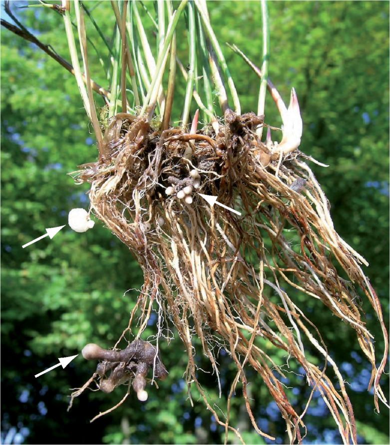
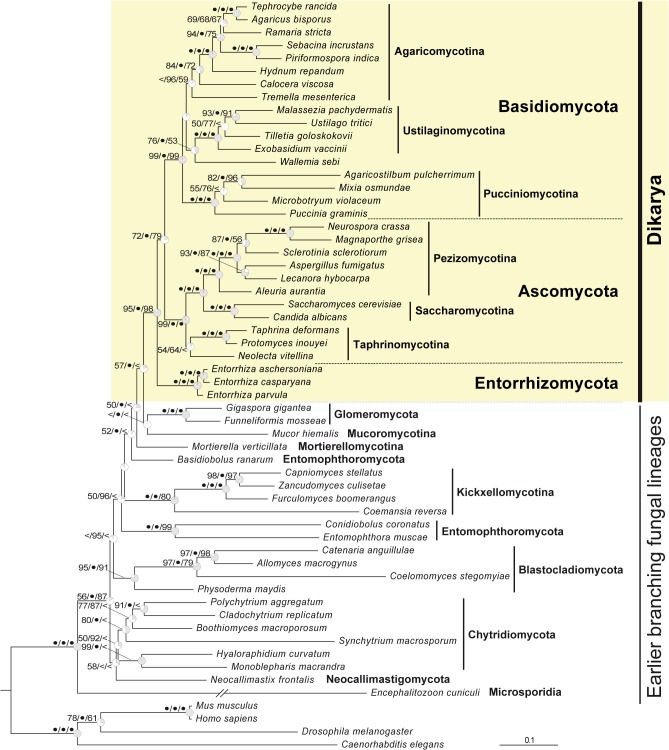
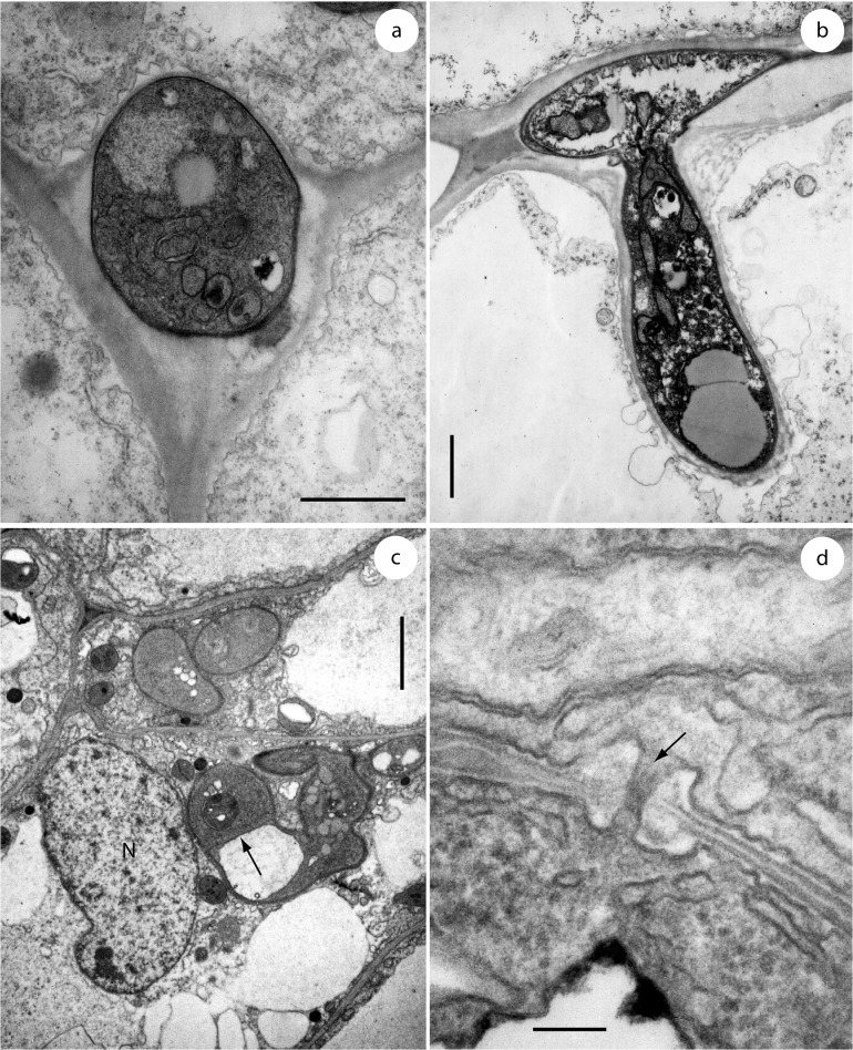
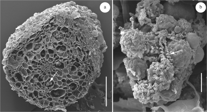
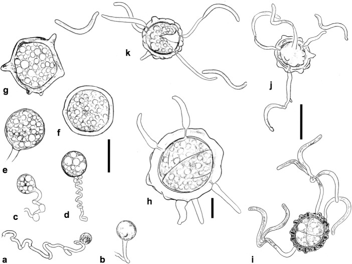
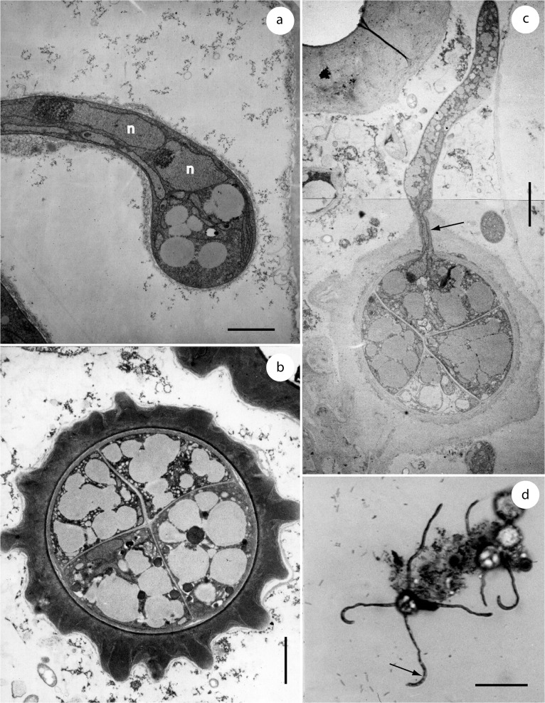
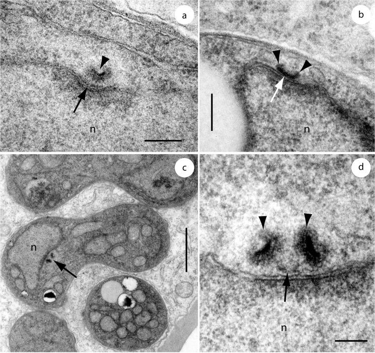
Entorrhiza is a small fungal genus comprising 14 species that all cause galls on roots of Cyperaceae and Juncaceae. Although this genus was established 130 years ago, crucial questions on the phylogenetic relationships and biology of this enigmatic taxon are still unanswered. In order to infer a robust hypothesis about the phylogenetic position of Entorrhiza and to evaluate evolutionary trends, multiple gene sequences and morphological characteristics of Entorrhiza were analyzed and compared with respective findings in Fungi. In our comprehensive five-gene analyses Entorrhiza appeared as a highly supported monophyletic lineage representing the sister group to the rest of the Dikarya, a phylogenetic placement that received but moderate maximum likelihood and maximum parsimony bootstrap support. An alternative maximum likelihood tree with the constraint that Entorrhiza forms a monophyletic group with Basidiomycota could not be rejected. According to the first phylogenetic hypothesis, the teliospore tetrads of Entorrhiza represent the prototype of the dikaryan meiosporangium. The alternative hypothesis is supported by similarities in septal pore structure, cell wall and spindle pole bodies. Based on the isolated phylogenetic position of Entorrhiza and its peculiar combination of features related to ultrastructure and reproduction mode, we propose a new phylum Entorrhizomycota, for the genus Entorrhiza, which represents an apparently widespread group of inconspicuous fungi.


Conflict of interest statement

Competing Interests: The authors have declared that no competing interests exist.

Figures

Fig 1
Fig 1. Portion of a tussock of Juncus articulatus with root galls caused by Entorrhiza casparyana (arrows).
Note that the older segments of the galls are brown-colored while younger parts are whitish (tips).
Fig 2
Fig 2. Maximum likelihood tree showing the phylogenetic position of the genus Entorrhiza within the kingdom Fungi derived from a combined dataset of rDNA (18S, 5.8S and 28S) and amino acid (RPB1 and RPB2) sequences.
Branch support is given as maximum likelihood bootstrap percentage (1,000 replicates) / Bayesian posterior probability (consensus of two independent MCMC processes, each with four chains over five million generations) / maximum parsimony bootstrap (1,000 replicates). Values of 100% are designated with bullets, values below 50% are omitted or designated by <. Grey sectors in the clock symbols illustrate internode confidence values (ICA [30]) of the supporting branch; counterclockwise orientation designates a negative ICA value, i.e., an instance where a conflicting bipartition occurs more frequently in the bootstrap trees than the bipartition defined by this branch. The tree was rooted with the Metazoa branch. The Encephalitozoon branch was scaled with a factor of 0.5 for graphical reasons. For GenBank accession numbers see Table 1.
Fig 3
Fig 3. Soral hyphae of Entorrhiza casparyana in root galls of Juncus articulatus, as seen by TEM.
(a) Section through an intercellular hypha surrounded by plant cells. (b) Section through an intercellular hypha extended into a cortical cell. Note that the intracellular part of the hypha is surrounded by the host plasma membrane. (c) Section through two hyphal coils in neighbouring cortical cells of J. articulatus. One host nucleus is visible at N, and a hyphal septum is visible at the arrow. (d) Section through a hyphal septum showing a dolipore without membranous caps (arrow). Note the basidiomycetous tripartite profile of the septum wall. Scale bar is 1 μm in (a-b), 2 μm in (c), and 0.1 μm in (d).
Fig 4
Fig 4. Sections through galls of Entorrhiza casparyana on Juncus articulatus, as seen by SEM.
(a) Section showing intracellular teliospore packets (one is indicated by an arrow). (b) Teliospore packet in detail, showing teliospores and hyphal spirals (one is indicated by an arrow). Scale bar is 1 mm in (a) and 20 μm in (b).
Fig 5
Fig 5. Line drawings of Entorrhiza casparyana.
(a-g) Intracellular more or less coiled hyphae and teliospores in different developing stages (host cells not drawn). (h-k) Germinating teliospores in different developing stages. Note the sigmoid shape of the propagules. Scale bar is 20 μm in (a-g, j-k) and 10 μm in (h).
Fig 6
Fig 6. Teliospore formation and germination of Entorrhiza casparyana, as seen by TEM (a-c) or LM (d).
(a) Section through an intracellular hyphal coil showing a terminal teliospore initial with two nuclei (n). (b) Section through a teliospore tetrad showing the cruciform septation. (c) Section through a teliospore tetrad showing the germination of one of the four teliospore compartments. One nucleus is visible at the arrow, probably migrating into the germination hypha. (d) Light microscopic micrograph of a germinating teliospore illustrated to show the synchronous development of the germination hyphae and the terminal sigmoid propagules (one is indicated by an arrow). Scale bar is 2 μm in (a-b), 3 μm in (c), and 40 μm in (d).
Fig 7
Fig 7. Spindle pole bodies (SPBs) of Entorrhiza casparyana, as seen by TEM.
(a) Median section through a postmitotic / early interphasic SPB showing the original hemispherical SPB with an internal electron-opaque layer (arrowhead) and an additional bar (arrowhead) next to the nuclear envelope. (b) Longitudinal section through a mid-interphasic SPB. The double structure of the SPB has begun to reconstitute by growth of SPB elements (arrowheads) at the ends of the electron-opaque bar (arrow). (c) Section through an intracellular hyphal coil showing one nucleus with a longitudinally sectioned premitotic SPB (arrow). (d) Detail of (c) illustrated to show the two enlarged SPB elements (arrowheads) connected by an elongated middle piece (arrow). Note the internal layering of the SPB elements. n, nucleus. Scale bar is 0.2 μm in (a-b), 1 μm in (c), and 0.1 μm in (d).

Similar articles

Cited by

References

    1. Weber C (1884) Über den Pilz der Wurzelanschwellungen von Juncus bufonius . Botanische Zeitung 42: 369–379.
    1. Vánky K (2012) Smut Fungi of the World. St. Paul: APS.
    1. Fineran JM (1980) The structure of galls induced by Entorrhiza C. Weber (Ustilaginales) on roots of the Cyperaceae and Juncaceae. Nova Hedwigia 32: 265–284.
    1. Deml G, Oberwinkler F (1981) Investigations on Entorrhiza casparyana by light and electron microscopy. Mycologia 73: 392–398.
    1. Fineran JM (1978) A taxonomic revision of the genus Entorrhiza C. Weber (Ustilaginales). Nova Hedwigia 30: 1–67.

Publication types

LinkOut - more resources