

Ultrasound in Radiology: From Anatomic, Functional, Molecular Imaging to Drug Delivery and Image-Guided Therapy
- PMID: 26200224
- PMCID: PMC4580624
- DOI: 10.1097/RLI.0000000000000188
Ultrasound in Radiology: From Anatomic, Functional, Molecular Imaging to Drug Delivery and Image-Guided Therapy
Abstract
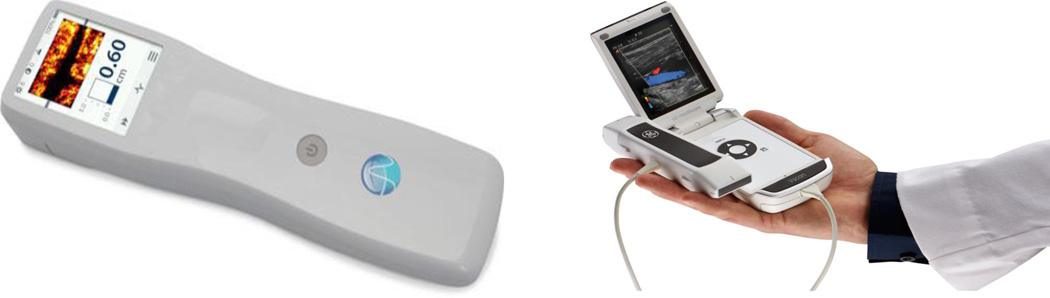
During the past decade, ultrasound has expanded medical imaging well beyond the "traditional" radiology setting: a combination of portability, low cost, and ease of use makes ultrasound imaging an indispensable tool for radiologists as well as for other medical professionals who need to obtain imaging diagnosis or guide a therapeutic intervention quickly and efficiently. Ultrasound combines excellent ability for deep penetration into soft tissues with very good spatial resolution, with only a few exceptions (ie, those involving overlying bone or gas). Real-time imaging (up to hundreds and thousands of frames per second) enables guidance of therapeutic procedures and biopsies; characterization of the mechanical properties of the tissues greatly aids with the accuracy of the procedures. The ability of ultrasound to deposit energy locally brings about the potential for localized intervention encompassing the following: tissue ablation, enhancing penetration through the natural barriers to drug delivery in the body and triggering drug release from carrier microparticles and nanoparticles. The use of microbubble contrast agents brings the ability to monitor and quantify tissue perfusion, and microbubble targeting with ligand-decorated microbubbles brings the ability to obtain molecular biomarker information, that is, ultrasound molecular imaging. Overall, ultrasound has become the most widely used imaging modality in modern medicine; it will continue to grow and expand.
Figures
Similar articles
-
From Anatomy to Functional and Molecular Biomarker Imaging and Therapy: Ultrasound Is Safe, Ultrafast, Portable, and Inexpensive.Invest Radiol. 2020 Sep;55(9):559-572. doi: 10.1097/RLI.0000000000000675. Invest Radiol. 2020. PMID: 32776766 Free PMC article. Review.
-
[Progress of ultrasound microbubble contrast technology in the diagnosis and treatment of clinical diseases].Sichuan Da Xue Xue Bao Yi Xue Ban. 2014 Nov;45(6):974-8. Sichuan Da Xue Xue Bao Yi Xue Ban. 2014. PMID: 25571727 Chinese.
-
Perfusion-guided sonopermeation of neuroblastoma: a novel strategy for monitoring and predicting liposomal doxorubicin uptake in vivo.Theranostics. 2020 Jul 9;10(18):8143-8161. doi: 10.7150/thno.45903. eCollection 2020. Theranostics. 2020. PMID: 32724463 Free PMC article.
-
The role of ultrasound in molecular imaging.Br J Radiol. 2003;76 Spec No 2:S140-50. doi: 10.1259/bjr/57063872. Br J Radiol. 2003. PMID: 15572336 Review.
-
Microbubble-mediated intravascular ultrasound imaging and drug delivery.IEEE Trans Ultrason Ferroelectr Freq Control. 2015 Sep;62(9):1674-85. doi: 10.1109/TUFFC.2015.007143. IEEE Trans Ultrason Ferroelectr Freq Control. 2015. PMID: 26415129
Cited by
-
Improved Sensitivity in Ultrasound Molecular Imaging With Coherence-Based Beamforming.IEEE Trans Med Imaging. 2018 Jan;37(1):241-250. doi: 10.1109/TMI.2017.2774814. IEEE Trans Med Imaging. 2018. PMID: 29293430 Free PMC article.
-
Ultrasound-Triggered Amoxicillin Release from Chitosan/Ethylene Glycol Diglycidyl Ether/Amoxicillin Hydrogels Having a Covalently Bonded Network.ACS Omega. 2023 Dec 19;9(1):585-597. doi: 10.1021/acsomega.3c06213. eCollection 2024 Jan 9. ACS Omega. 2023. PMID: 38222581 Free PMC article.
-
Normology: Is it Time to Rethink Point-of-Care Ultrasound Training?J Med Educ Curric Dev. 2024 Feb 12;11:23821205241232498. doi: 10.1177/23821205241232498. eCollection 2024 Jan-Dec. J Med Educ Curric Dev. 2024. PMID: 38357688 Free PMC article.
-
Comparison of ultrasound- vs. landmark-guided injections for musculoskeletal pain: an umbrella review.J Rehabil Med. 2024 Aug 26;56:jrm40679. doi: 10.2340/jrm.v56.40769. J Rehabil Med. 2024. PMID: 39185547 Free PMC article.
-
Ultrasound-mediated mechanical forces activate selective tumor cell apoptosis.Bioeng Transl Med. 2024 Dec 30;10(2):e10737. doi: 10.1002/btm2.10737. eCollection 2025 Mar. Bioeng Transl Med. 2024. PMID: 40060768 Free PMC article.
References
-
- Szabo T. ISBN: 9780123964878. 2nd Edition. Elsevier; 2013. Diagnostic Ultrasound Imaging: Inside Out.
-
- Moore G. Cramming More Components onto Integrated Circuits. Electronics. 1965:114–117.
-
- Tanter M, Fink M. Ultrafast Imaging in Biomedical Ultrasound. IEEE Transactions on Ultrasonics Ferroelectrics and Frequency Control. 2014;61:102–119. - PubMed
-
- Schneider FK, Agarwal A, Yoo YM, et al. A Fully Programmable Computing Architecture for Medical Ultrasound Machines. Ieee Transactions on Information Technology in Biomedicine. 2010;14:538–540. - PubMed
-
- Blaivas M, Brannam L, Theodoro D. Ultrasound image quality comparison between an inexpensive handheld emergency department (ED) ultrasound machine and a large mobile ED ultrasound system. Academic Emergency Medicine. 2004;11:778–781. - PubMed
Publication types
MeSH terms
Grants and funding
- EB002349/EB/NIBIB NIH HHS/United States
- R01 EB001826/EB/NIBIB NIH HHS/United States
- R01 HL090700/HL/NHLBI NIH HHS/United States
- EB001826/EB/NIBIB NIH HHS/United States
- R21 DK093841/DK/NIDDK NIH HHS/United States
- DK093841/DK/NIDDK NIH HHS/United States
- EB016752/EB/NIBIB NIH HHS/United States
- R21 EB016752/EB/NIBIB NIH HHS/United States
- HL111077/HL/NHLBI NIH HHS/United States
- CA102880/CA/NCI NIH HHS/United States
- HL090700/HL/NHLBI NIH HHS/United States
- R01 EB002349/EB/NIBIB NIH HHS/United States
- R21 CA102880/CA/NCI NIH HHS/United States
- R33 CA102880/CA/NCI NIH HHS/United States
- R01 HL111077/HL/NHLBI NIH HHS/United States
LinkOut - more resources
Full Text Sources
Miscellaneous
