. 2015 Jul 22;10(7):e0132867.
doi: 10.1371/journal.pone.0132867. eCollection 2015.

The Glycogen Synthase Kinase 3α and β Isoforms Differentially Regulates Interleukin-12p40 Expression in Endothelial Cells Stimulated with Peptidoglycan from Staphylococcus aureus

Affiliations

The Glycogen Synthase Kinase 3α and β Isoforms Differentially Regulates Interleukin-12p40 Expression in Endothelial Cells Stimulated with Peptidoglycan from Staphylococcus aureus

Ricarda Cortés-Vieyra et al. PLoS One. .

Abstract

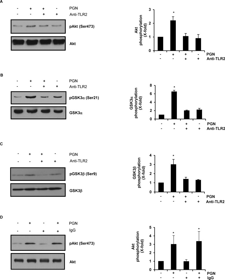
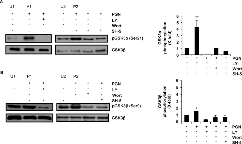
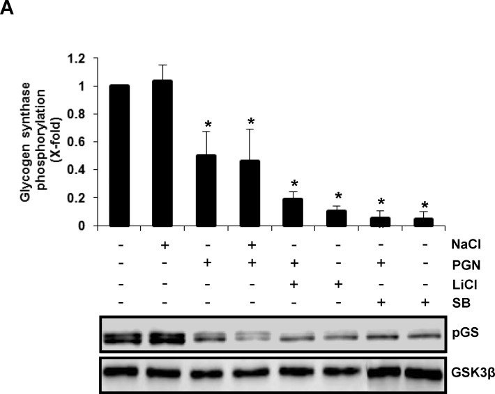
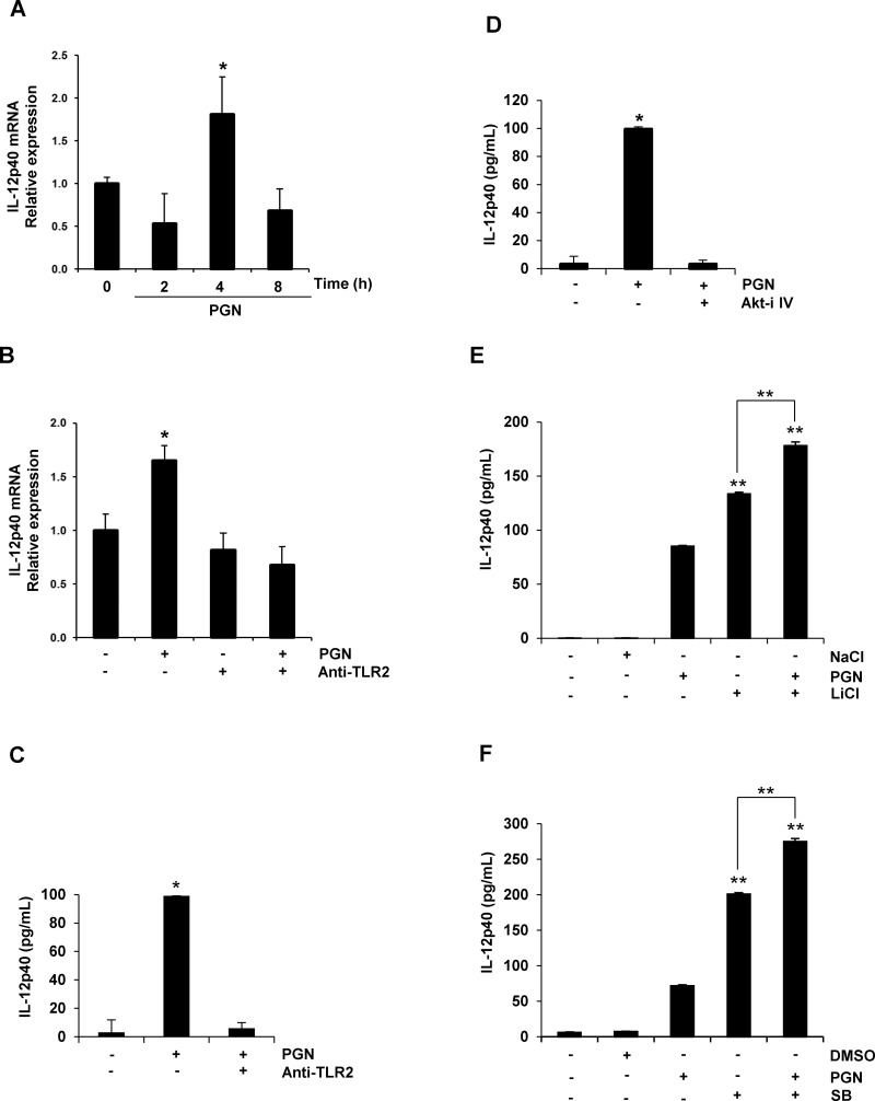
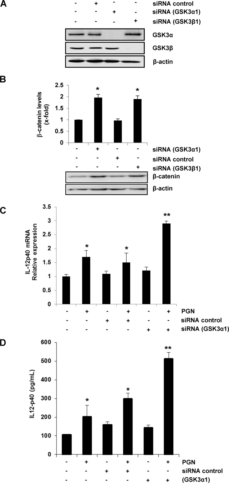
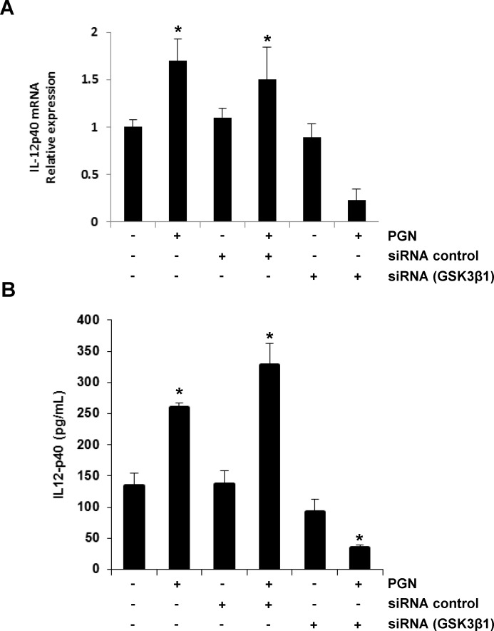
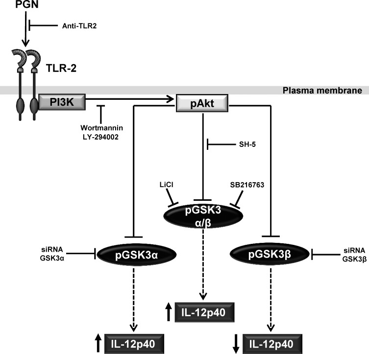
Glycogen synthase kinase 3 (GSK3) is a constitutively active regulatory enzyme that is important in cancer, diabetes, and cardiovascular, neurodegenerative, and psychiatric diseases. While GSK3α is usually important in neurodegenerative and psychiatric diseases GSK3β is fundamental in the inflammatory response caused by bacterial components. Peptidoglycan (PGN), one of the most abundant cell-wall structures of Gram-positive bacteria, is an important inducer of inflammation. To evaluate whether inhibition of GSK3α and GSK3β activity in bovine endothelial cells (BEC) regulates the expression of the pro-inflammatory cytokine IL-12p40, we treated BEC with SDS-purified PGN from Staphylococcus aureus. We found that PGN triggered a TLR2/PI3K/Akt-dependent phosphorylation of GSK3α at Ser21, GSK3β at Ser9, and NF-κB p65 subunit (p65) at Ser536, and the phosphorylation of GSK3α was consistently higher than that of GSK3β. The expression of IL-12p40 was inhibited in BEC stimulated with PGN and pre-treated with a specific neutralizing anti-TLR2 antibody that targets the extracellular domain of TLR2 or by the addition of Akt-i IV (an Akt inhibitor). Inhibition of GSK3α and GSK3β with LiCl or SB216763 induced an increase in IL-12p40 mRNA and protein. The effect of each isoform on IL-12p40 expression was evaluated by siRNA-gene expression silencing of GSK3α and GSK3β. GSK3α gene silencing resulted in a marked increase in IL-12p40 mRNA and protein while GSK3β gene silencing had the opposite effect on IL-12p40 expression. These results indicate that the TLR2/PI3K/Akt-dependent inhibition of GSK3α activity also plays an important role in the inflammatory response caused by stimulation of BEC with PGN from S. aureus.


Conflict of interest statement

Competing Interests: The authors have declared that no competing interests exist.

Figures

Fig 1
Fig 1. Peptidoglycan (PGN) induces phosphorylation of Akt, GSK3α and GSK3β in bovine endothelial cells (BEC) through TLR2 activation.
A, B, C) BEC were either stimulated with 10 μg/mL of PGN for 30 min, pre-incubated with 5 μg/mL of neutralizing antibody against the extracellular domain of TLR2 (anti-TLR2) for 60 min and stimulated with 10 μg/mL of PGN for 30 min or pre-incubated with 5 μg/mL of anti-TLR2 for 60 min. D) BEC were either stimulated with 10 μg/mL of PGN for 30 min, pre-incubated with 5 μg/mL of an isotype anti-IgG for 60 min or pre-incubated with 5 μg/mL of anti-IgG for 60 min and stimulated with 10 μg/mL of PGN for 30 min. As control in all cases A-D, BEC were left untreated (-). After treatments, protein extracts were analyzed by western blot and probed with monoclonal antibodies against the phosphorylated forms of Akt1 (pAkt Ser473), GSK3α (pGSK3α Ser21) or GSK3β (pGSK3β Ser9). Blots were stripped and reprobed with an antibody that recognizes the nonphosphorylated forms of Akt (A, and D), GSK3α (B) or GSK3β (C) to verify equal protein loading. Blots are representative of three independent experiments. Graphs on the right panel indicate the band intensity obtained by densitometric analysis. Results are expressed as the mean ± S.E.M. (n = 3). *p < 0.05, compared with the untreated control.
Fig 2
Fig 2. PGN induces PI3K/Akt-dependent phosphorylation of GSK3α and GSK3β in BEC.
A-B) BEC were untreated and unstimulated (U1 and U2), stimulated with 10 μg/mL of PGN for 30 min (P1 and P2) or treated with either 10 μM of LY294002 (LY), 100 nM of Wortmannin (Wort) or 10 μM of SH-5 for 30 min, and then stimulated with 10 μg/mL of PGN for 30 min. Protein extracts were analyzed by western blot and probed with monoclonal antibodies against the phosphorylated forms of GSK3α (pGSK3α Ser21) or GSK3β (pGSK3β Ser9. To verify for equal amount of proteins, blots were stripped and reprobed with an antibody that recognizes the nonphosphorylated form of GSK3β. Blots are representative of three independent experiments. Graphs indicate the band intensity obtained by densitometric analysis. In each graph, the densitometric control values plotted were the average of U1 + U2 while the values plotted for the PGN-stimulated cells were the average of P1 + P2. Results are expressed as the mean ± S.E.M. (n = 3). *p < 0.05; **p < 0.01, compared with the unstimulated control.
Fig 3
Fig 3. Inhibition of glycogen synthase phosphorylation at Ser641 in BEC treated with PGN, LiCl or SB, and stabilization of β-catenin levels by GSK3α or GSK3β gene silencing.
A) BEC were either stimulated with 10 μg/mL of PGN for 30 min, treated with 10 mM NaCl for 60 min, 10 mM LiCl or 10 μM SB and then stimulated with 10 μg/mL of PGN for 30 min. As controls BEC were left untreated (-) or incubated with NaCl, LiCl or SB alone for 60 min. B) BEC were transfected with siRNA targeting GSK3α (siRNA GSK3α), siRNA targeting GSK3β (siRNA GSK3β), control siRNA (siRNA control) or left untransfected (-) for 16 days. After transfection, BEC were incubated for 24 h. Protein extracts were analyzed by western blot and probed with a monoclonal antibody against the phosphorylated form of glycogen synthase (GS Ser641) or the nonphosphorylated form of β-catenin. To verify that equal amount of protein was loaded in each lane, blots were stripped and reprobed with antibodies that recognize the nonphosphorylated forms of GSK3β (A) or β-actin (B). Blots are representative of three independent experiments. Graphs indicate the band intensity obtained by densitometric analysis. Results are expressed as the mean ± S.E.M. (n = 3). *p<0.05, compared with the untreated (A) or untransfected (B) control.
Fig 4
Fig 4. PGN induces IL-12p40 expression through TLR2/Akt activation and GSK3 inhibition in BEC.
A) BEC were left untreated and unstimulated (0) or stimulated with 10 μg/mL of PGN-prep for 2, 4 or 8 h. B) BEC were stimulated with 10 μg/mL of PGN for 4 h, treated with 5 μg/mL of neutralizing anti-TLR2 for 60 min, or treated with 5 μg/mL of anti-TLR2 for 60 min and then stimulated with 10 μg/mL of PGN for 4 h. C) BEC were stimulated with 10 μg/mL of PGN for 9 h or treated with 5 μg/mL of anti-TLR2 for 60 min and then stimulated with 10 μg/mL of PGN for 9 h. D) BEC were stimulated with 10 μg/mL of PGN for 9 h or pretreated with 1 μM of Akt inhibitor IV (Akt-i IV) for 30 min and then stimulated with 10 μg/mL for 9 h. E) BEC were treated with 10 mM NaCl for 60 min, stimulated with 10 μg/mL of PGN for 9 h, treated with 10 mM of LiCl for 60 min or treated with 10 mM of LiCl for 60 min and then stimulated with 10 μg/mL of PGN for 9 h. F) BEC were treated with 10 μM of DMSO, stimulated with 10 μg/mL of PGN for 9 h, treated with 10 μM of SB 216763 (SB) for 30 min or treated with 10 μM of SB 216763 (SB) for 30 min and then stimulated with 10 μg/mL of PGN for 9 h. As controls, in B-F BEC were left untreated and stimulated (-). Total RNA was extracted and relative transcript level of IL-12p40 was quantitated by qRT-PCR using the delta-delta Ct method, and amplification of β-actin as a reference gene (A-B). Cell-free supernatants were analyzed by ELISA for production of IL-12p40 (C-F). Results are expressed as the mean ± S.E.M. (n = 3). *p <0.05; **p <0.01. All data were compared with the untreated and unstimulated controls.
Fig 5
Fig 5. GSK3α inhibition up-regulates IL-12p40 expression in BEC stimulated with PGN.
A) BEC were left untransfected (-), transfected with control siRNA (siRNA control), transfected with siRNA targeting GSK3α (siRNA GSK3α1) or transfected with siRNA targeting GSK3β (siRNA GSK3β1). B) BEC were left untransfected and unstimulated (-), stimulated with 10 μg/mL of PGN for 4 h, transfected with control siRNA (siRNA control), transfected with control siRNA and then stimulated with 10 μg/mL PGN for 4 h, transfected with siRNA targeting GSK3α (siRNA GSK3α1) or transfected with siRNA targeting GSK3α (siRNA GSK3α1) and then stimulated with 10 μg/mL of PGN for 4 h. C) BEC were left untransfected and unstimulated (-), stimulated with 10 μg/mL of PGN for 9 h, transfected with control siRNA (siRNA control), transfected with control siRNA and then stimulated with 10 μg/mL of PGN for 9 h, transfected with siRNA targeting GSK3α (siRNA GSK3α1) or transfected with siRNA targeting GSK3α1 and then stimulated with 10 μg/mL of PGN for 9 h. Protein extracts were analyzed by western blot and probed with a monoclonal antibody against the phosphorylated forms of GSK3α and GSK3β (A). To verify that equal amount of proteins was loaded in each lane, blots were stripped and reprobed with an antibody that recognizes the nonphosphorylated form of β-actin (A). Blots are representative of three independent experiments. Total RNA was extracted and relative transcript level of IL-12p40 was quantitated by qRT-PCR, using the delta-delta Ct method and amplification of β-actin as a reference gene (B). Cell-free supernatants were analyzed by ELISA for production of IL-12p40 (C). Results are expressed as the mean ± S.E.M. (n = 3). *p <0.05; **p <0.01. All data were compared with the untreated and untransfected controls.
Fig 6
Fig 6. GSK3β inhibition down-regulates IL-12p40 expression in BEC stimulated with PGN.
A) BEC were left untransfected and unstimulated (-), stimulated with 10 μg/mL of PGN for 4 h, transfected with control siRNA (siRNA control), transfected with control siRNA and then stimulated with 10 μg/mL of PGN for 4 h, transfected with siRNA targeting GSK3β (siRNA GSK3β1) or transfected with siRNA targeting GSK3β (siRNA GSK3β1) and then stimulated with 10 μg/mL of PGN for 4 h. B) BEC were left untransfected and unstimulated (-), stimulated with 10 μg/mL of PGN for 9 h, transfected with control siRNA (siRNA control), transfected with control siRNA and then stimulated with 10 μg/mL of PGN for 9 h, transfected with siRNA targeting GSK3β (siRNA GSK3β1) or transfected with siRNA targeting GSK3β (siRNA GSK3β1) and then stimulated with 10 μg/mL of PGN for 9 h. Total RNA was extracted and relative transcript level of IL-12p40 was quantitated by qRT-PCR, using the delta-delta Ct method, and amplification of β-actin as a reference gene (A). Cell-free supernatants were analyzed by ELISA for production of IL-12p40 (B). Results are expressed as the mean ± S.E.M. (n = 3). *p <0.05. All data were compared with the untreated and untransfected controls.
Fig 7
Fig 7. A working model of the signaling pathway involved on the IL-12p40 expression induced by PGN in BEC.
PGN induces the activation of the TLR2/PI3K/Akt pathway, which in turn induces GSK3α and GSK3β phosphorylation/inhibition and this in turn up- or down-regulates IL-12p40 expression. Anti-TLR2, Wortmannin (PI3K inhibitor), LY-294002 (PI3K inhibitor) or SH-5 (Akt inhibitor) blocks the phosphorylation/inhibition of GSK3α and GSK3β. LiCl (GSK3 inhibitor), SB-216763 (GSK3 inhibitor) or siRNA GSK3α increases the expression of IL-12p40 while siRNA GSK3β decreases the expression of IL-12p40. Not represented in the diagram is the decrease in IL-12p40 expression when TLR2 is blocked with anti-TLR2 and when Akt is inhibited with Akt-i IV.

Similar articles

Cited by

References

    1. Pantosti A. Methicillin-resistant Staphylococcus aureus associated with animals and its relevance to human health. Front Microbiol. 2012;3: 127 10.3389/fmicb.2012.00127 - DOI - PMC - PubMed
    1. McDonald C, Inohara N, Núñez G. Peptidoglycan signaling in innate immunity and inflammatory disease. J Biol Chem. 2005;280: 20177–20180. - PubMed
    1. Chen BC, Kang JC, Lu YT, Hsu MJ, Liao CC, Chiu WT, et al. Rac1 regulates peptidoglycan-induced nuclear factor-kappaB activation and cyclooxygenase-2 expression in RAW 264.7 macrophages by activating the phosphatidylinositol 3-kinase/Akt pathway. Mol Immunol. 2009;46: 1179–1188. 10.1016/j.molimm.2008.11.006 - DOI - PubMed
    1. Lin HY, Tang CH, Chen YH, Wei IH, Chen JH, Lai CH, et al. Peptidoglycan enhances proinflammatory cytokine expression through the TLR2 receptor, MyD88, phosphatidylinositol 3-kinase/AKT and NF-kappaB pathways in BV-2 microglia. Int Immunopharmacol. 2010;10: 883–891. 10.1016/j.intimp.2010.04.026 - DOI - PubMed
    1. Chiu YC, Lin CY, Chen CP, Huang KC, Tong KM, Tzeng CY, et al. Peptidoglycan enhances IL-6 production in human synovial fibroblasts via TLR2 receptor, focal adhesion kinase, Akt, and AP-1- dependent pathway. J Immunol. 2009;183: 2785–2792. 10.4049/jimmunol.0802826 - DOI - PubMed

Publication types

MeSH terms