. 2015 Jul;56(8):4560-9.
doi: 10.1167/iovs.15-16378.

Sarpogrelate, a 5-HT2A Receptor Antagonist, Protects the Retina From Light-Induced Retinopathy

Affiliations

Sarpogrelate, a 5-HT2A Receptor Antagonist, Protects the Retina From Light-Induced Retinopathy

Brandon E Tullis et al. Invest Ophthalmol Vis Sci. 2015 Jul.

Abstract

Purpose: To determine if sarpogrelate, a selective 5-HT2A receptor antagonist, is protective against light-induced retinopathy in BALB/c mice.

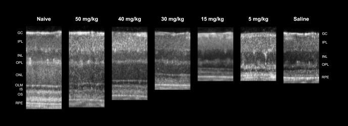
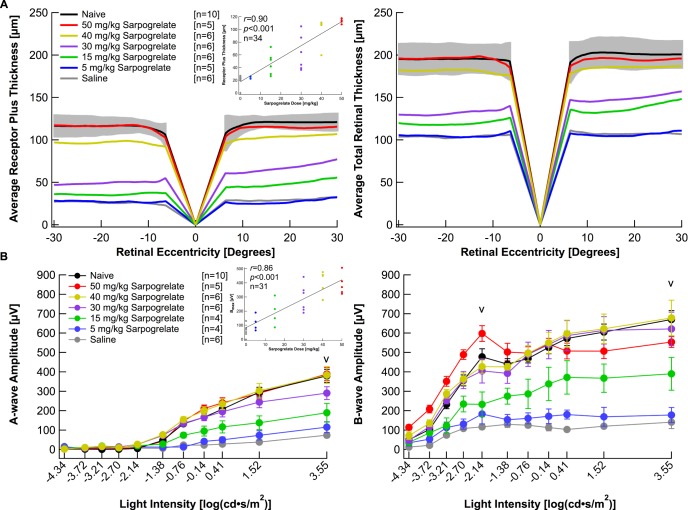
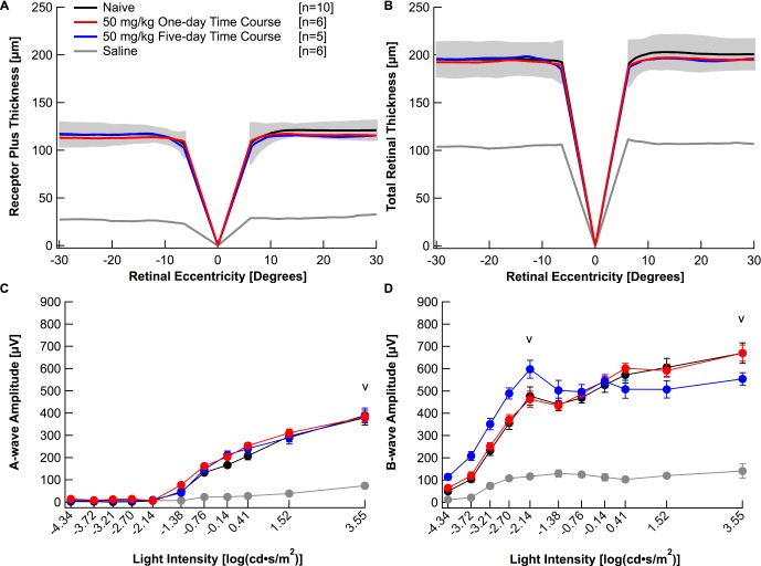
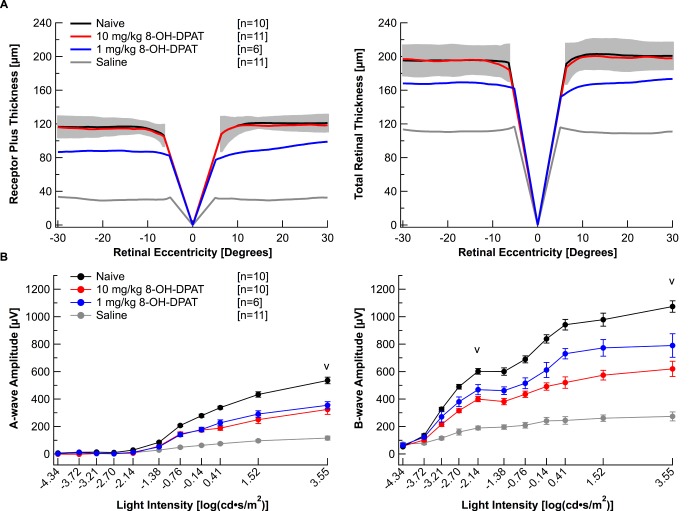
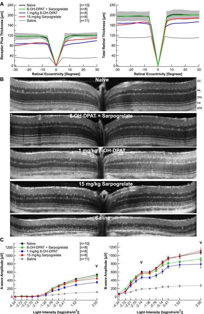
Methods: BALB/c mice were dosed intraperitoneally with 5, 15, 30, 40, or 50 mg/kg sarpogrelate 48, 24, and 0 hours prior to bright light exposure (10,000 lux) as well as 24 and 48 hours after exposure. Additionally, a single injection regimen was evaluated by injecting mice with 50 mg/kg sarpogrelate once immediately prior to light exposure. To investigate the potential for additive effects of serotonin receptor agents, a combination therapy consisting of sarpogrelate (15 mg/kg) and 8-OH-DPAT (1 mg/kg) was evaluated with the 5-day treatment regimen. Neuroprotection was characterized by the preservation of retinal thickness and function, measured by spectral-domain optical coherence tomography (SD-OCT) and electroretinography (ERG), respectively.

Results: Mice that were light damaged and injected with saline had significantly reduced outer retinal thickness, total retinal thickness, and ERG amplitudes compared with naïve mice. A 5-day administration of 15, 30, or 40 mg/kg of sarpogrelate was able to partially protect retinal morphology and full protection of retinal morphology was achieved with a 50 mg/kg dose. Both 15 and 30 mg/kg doses of sarpogrelate partially preserved retinal function measured by ERG, whereas 40 and 50 mg/kg doses fully preserved retinal function. Additionally, a single administration of 50 mg/kg sarpogrelate was able to fully preserve both retinal morphology and function. Administration of 15 mg/kg of sarpogrelate and 1 mg/kg of 8-OH-DPAT together demonstrated an additive effect and fully preserved retinal morphology.

Conclusions: A 5- or 1-day treatment with 50 mg/kg sarpogrelate can completely protect the retina of BALB/c mice from light-induced retinopathy. Partial protection can be achieved with lower doses starting at 15 mg/kg and protection increases in a dose-dependent manner. Treatment with low doses of sarpogrelate and 8-OH-DPAT elicits an additive effect that results in full protection of retinal morphology.


Figures

Figure 1
Figure 1
Sarpogrelate protects retinal morphology in a dose-dependent manner. Representative linear SD-OCT scans of nasal retina from a naïve mouse or mice injected with either 50, 40, 30, 15, or 5 mg/kg of sarpogrelate or saline. Injections followed the 5-day time course with a 6-hour light damage.
Figure 2
Figure 2
Sarpogrelate elicits neuroprotective effects in a dose-dependent manner. (A) Receptor plus thicknesses and total retinal thicknesses obtained from segmented horizontal SD-OCT scans were averaged and plotted versus retinal eccentricity from the optic nerve head. The gray area indicates ±2 SD of the naïve averaged data. The correlation coefficient, r, was determined by comparing right eye REC+ thicknesses to the dose of sarpogrelate administered. (B) Electroretinogram a- and b-wave amplitudes from mice in each group were extracted from individual waveforms, averaged and plotted versus the light intensity of each flash. Averages are represented as mean ± standard error. “V” indicates at which light intensity the statistical analysis was performed. The correlation coefficient, r, was determined by comparing each animal's amax amplitude to the dose of sarpogrelate administered. Three animals died after acquiring OCT images, but prior to ERG recordings. Injections followed the 5-day time course with a 6-hour light damage. Black: Naïve. Red: 50 mg/kg sarpogrelate. Yellow: 40 mg/kg sarpogrelate. Purple: 30 mg/kg sarpogrelate. Green: 15 mg/kg sarpogrelate. Blue: 5 mg/kg sarpogrelate. Gray: Saline.
Figure 3
Figure 3
A 1-day time course of 50 mg/kg sarpogrelate completely protects retinal morphology and function. Receptor plus thicknesses (A) and total retinal thicknesses (B) obtained from segmented horizontal SD-OCT scans were averaged and plotted versus retinal eccentricity from the optic nerve head. The gray area indicates ±2 SD of the naïve averaged data. Electroretinogram a- (C) and b-wave (D) amplitudes for all mice in each group were extracted from individual waveforms, averaged and plotted versus the light intensity of each flash. Averages are represented as mean ± standard error. “V” indicates at which light intensity the statistical analysis was performed. Black: Naïve. Red: 50 mg/kg 1-day time course. Blue: 50 mg/kg 5-day time course. Gray: Saline.
Figure 4
Figure 4
Chemical 8-OH-DPAT elicits neuroprotective effects in BALB/c mice. (A) Receptor plus thicknesses and total retinal thicknesses obtained from segmented horizontal SD-OCT scans were averaged and plotted versus retinal eccentricity from the optic nerve head. The gray area indicates ±2 SD of the naïve averaged data. (B) Electroretinogram a- and b-wave amplitudes from mice in each group were extracted from individual waveforms, averaged, and plotted versus the light intensity of each flash. Averages are represented as mean ± standard error. “V” indicates at which light intensity the statistical analysis was performed. One animal died after acquiring OCT images, but prior to ERG recordings. Injections followed the 5-day time course with a 1-hour light damage. Black: Naïve. Red: 10 mg/kg 8-OH-DPAT. Blue: 1 mg/kg 8-OH-DPAT. Gray: Saline.
Figure 5
Figure 5
A combination therapy of sarpogrelate and 8-OH-DPAT provide additive neuroprotection. (A) Receptor plus thicknesses and total retinal thicknesses obtained from segmented horizontal SD-OCT scans were averaged and plotted versus retinal eccentricity from the optic nerve head. The gray area indicates ±2 SD of the naïve averaged data. (B) Representative linear SD-OCT scans. (C) Electroretinogram a- and b-wave amplitudes from mice in each group were extracted from individual waveforms, averaged and plotted versus the light intensity of each flash. Averages are represented as mean ± standard error. “V” indicates at which light intensity the statistical analysis was performed. Injections followed the 5-day time course with a 1-hour light damage. Black: Naïve. Green: 8-OH-DPAT + Sarpogrelate. Blue: 1 mg/kg 8-OH-DPAT. Red: 15 mg/kg Sarpogrelate. Gray: Saline.

References

    1. Ueki Y,, Wang J,, Chollangi S,, Ash J. STAT3 activation in photoreceptors by leukemia inhibitory factor is associated with protection from light damage. J Neurochem. 2008; 105: 784–796. - PubMed
    1. Zhao L,, Wang C,, Song D,, et al. Systemic administration of the antioxidant/iron chelator α-lipoic acid protects against light-induced photoreceptor degeneration in the mouse retina. Invest Ophthalmol Vis Sci. 2014; 55: 5979–5988. - PMC - PubMed
    1. Song D,, Song Y,, Hadziahmetovic M,, Zhong Y,, Dunaief JL. Systemic administration of the iron chelator deferiprone protects against light-induced photoreceptor degeneration in the mouse retina. Free Radic Biol Med. 2012; 53: 64–71. - PMC - PubMed
    1. Imai S,, Inokuchi Y,, Nakamura S,, Tsuruma K,, Shimazawa M,, Hara H. Systemic administration of a free radical scavenger, edaravone, protects against light-induced photoreceptor degeneration in the mouse retina. Eur J Pharmacol. 2010; 642: 77–85. - PubMed
    1. Razaie T,, McKercher SR,, Kosaka K,, et al. Protective effect of carnosic acid, a pro-electrophilic compound, in models of oxidative stress and light-induced retinal degeneration. Invest Ophthalmol Vis Sci. 2012; 53: 7847–7854. - PMC - PubMed

Publication types

MeSH terms

Substances