

Atypical Hepatocellular Neoplasm With Peliosis in Cirrhotic Liver Versus Hepatocellular Carcinoma: A Diagnostic Trap
- PMID: 26200629
- PMCID: PMC4603009
- DOI: 10.1097/MD.0000000000001189
Atypical Hepatocellular Neoplasm With Peliosis in Cirrhotic Liver Versus Hepatocellular Carcinoma: A Diagnostic Trap
Abstract
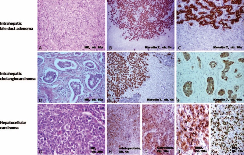
Atypical hepatocellular neoplasm (AHN) is an adenoma-like hepatic tumor that even occurs in noncirrhotic liver of males (any age) or females ≥ 50 years old, or associates focal atypical features. In this article, 2 unusual cases diagnosed in elderly cirrhotic patients, unrelated to steroids, are presented. The first case was incidentally diagnosed in an 83-year-old female. During laparoscopic surgery for cholecystectomy, hemoperitoneum was installed and laparotomy was necessary to remove a 70-mm nodular encapsulated hepatic tumor that was microscopically composed by hepatocyte-like cells with clear cytoplasm, arranged in 1- to 2-cell-thick plates and intermingled with areas of peliosis, negative for alpha fetoprotein (αFP), p53, and keratin 7, with low Ki67 index and intact reticulin framework. The second case was incidentally diagnosed at ultrasound examination in a 66-year-old male. The surgical specimen was a 50-mm solid multinodular tumor that microscopically consisted of 3-cell-thick plates of hepatocyte-like cells with acinar, pseudoglandular, and trabecular architecture, intermingled with peliotic areas, without nuclear atypia and disintegrated reticulin framework. Both of the cases occurred in cirrhotic liver. The tumor cells were marked by AE1/AE3 keratin, displayed a Ki67 index < 5% and were negative for αFP, p53, and keratin 7. No recurrences or any other disorder occurred 6 months after surgery. In cirrhotic liver, adenomas with peliosis that do not satisfy all the diagnosis criteria synthesized in the article should be considered AHNs and differential diagnosis includes hepatocellular carcinoma but also focal nodular hyperplasia, regenerative nodules, and dysplastic nodules. This histological entity is not yet included in the WHO Classification list.
Conflict of interest statement
The authors have no conflicts of interest to disclose.
Figures


Similar articles
-
Adenomatous hyperplastic nodules in the cirrhotic liver: differentiation from hepatocellular carcinoma with MR imaging.Radiology. 1989 Oct;173(1):123-6. doi: 10.1148/radiology.173.1.2550995. Radiology. 1989. PMID: 2550995
-
Giant hepatocellular adenoma with peliosis hepatis in a child: A diagnostic dilemma.Trop Gastroenterol. 2015 Jul-Sep;36(3):200-2. doi: 10.7869/tg.287. Trop Gastroenterol. 2015. PMID: 27522744 No abstract available.
-
[A rare form of benign tumor of the liver possibly related to the use of oral contraceptives: focal pediculated nodular hyperplasia].Rev Fr Gynecol Obstet. 1985 Aug-Sep;80(8-9):621-7. Rev Fr Gynecol Obstet. 1985. PMID: 2997901 French.
-
Problematic lesions in cirrhotic liver mimicking hepatocellular carcinoma.Eur Radiol. 2019 Sep;29(9):5101-5110. doi: 10.1007/s00330-019-06030-0. Epub 2019 Feb 20. Eur Radiol. 2019. PMID: 30788586 Review.
-
Differential diagnosis of hepatocellular nodular lesions.Semin Diagn Pathol. 1998 Nov;15(4):285-99. Semin Diagn Pathol. 1998. PMID: 9845429 Review.
Cited by
-
Synchronous Hepatocellular and Intrahepatic Cholangiocellular Carcinoma With Predominant Ductal Plate Malformation Pattern. A Case Report and Review of the Literature.Cancer Rep (Hoboken). 2025 Feb;8(2):e70085. doi: 10.1002/cnr2.70085. Cancer Rep (Hoboken). 2025. PMID: 39948704 Free PMC article. Review.
-
Combined hepatocellular-cholangiocarcinoma: from genesis to molecular pathways and therapeutic strategies.J Cancer Res Clin Oncol. 2024 May 23;150(5):270. doi: 10.1007/s00432-024-05781-8. J Cancer Res Clin Oncol. 2024. PMID: 38780656 Free PMC article. Review.
-
Multifocal nodular lesions in fatty liver mimicking neoplastic disease: a case report.Future Sci OA. 2023 Mar 28;9(4):FSO848. doi: 10.2144/fsoa-2022-0084. eCollection 2023 Apr. Future Sci OA. 2023. PMID: 37090491 Free PMC article.
-
Assessment of small hepatocellular carcinoma: perfusion quantification and time-concentration curve evaluation using color-coded and quantitative digital subtraction angiography.Medicine (Baltimore). 2018 Nov;97(48):e13392. doi: 10.1097/MD.0000000000013392. Medicine (Baltimore). 2018. PMID: 30508937 Free PMC article.
-
Serum microRNA panel for early diagnosis of the onset of hepatocellular carcinoma.Medicine (Baltimore). 2017 Jan;96(2):e5642. doi: 10.1097/MD.0000000000005642. Medicine (Baltimore). 2017. PMID: 28079796 Free PMC article.
References
-
- Turdean S, Gurzu S, Turcu M, et al. Current data in clinicopathological characteristics of primary hepatic tumors. Rom J Morphol Embryol 2012; 53:719–724. - PubMed
-
- Ferrell L, Kakar S. Fletcher DM, Christopher Tumor of the liver, gallbladder, and biliary tree. Diagnostic Histopathology of Tumors 3rd edChurchill Livingstone: Elsevier; 2007. 417–453.
Publication types
MeSH terms
LinkOut - more resources
Full Text Sources
Medical
Research Materials
Miscellaneous
