

Schistosome-induced cholangiocyte proliferation and osteopontin secretion correlate with fibrosis and portal hypertension in human and murine schistosomiasis mansoni
- PMID: 26201095
- PMCID: PMC4558314
- DOI: 10.1042/CS20150117
Schistosome-induced cholangiocyte proliferation and osteopontin secretion correlate with fibrosis and portal hypertension in human and murine schistosomiasis mansoni
Abstract
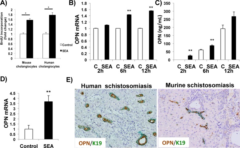
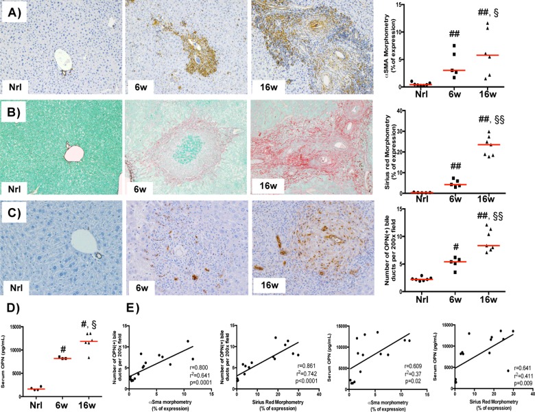
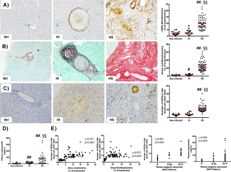
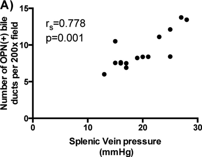
Schistosomiasis is a major cause of portal hypertension worldwide. It associates with portal fibrosis that develops during chronic infection. The mechanisms by which the pathogen evokes these host responses remain unclear. We evaluated the hypothesis that schistosome eggs release factors that directly stimulate liver cells to produce osteopontin (OPN), a pro-fibrogenic protein that stimulates hepatic stellate cells to become myofibroblasts. We also investigated the utility of OPN as a biomarker of fibrosis and/or severity of portal hypertension. Cultured cholangiocytes, Kupffer cells and hepatic stellate cells were treated with soluble egg antigen (SEA); OPN production was quantified by quantitative reverse transcriptase polymerase chain reaction (qRTPCR) and ELISA; cell proliferation was assessed by BrdU (5-bromo-2'-deoxyuridine). Mice were infected with Schistosoma mansoni for 6 or 16 weeks to cause early or advanced fibrosis. Liver OPN was evaluated by qRTPCR and immunohistochemistry (IHC) and correlated with liver fibrosis and serum OPN. Livers from patients with schistosomiasis mansoni (early fibrosis n=15; advanced fibrosis n=72) or healthy adults (n=22) were immunostained for OPN and fibrosis markers. Results were correlated with plasma OPN levels and splenic vein pressures. SEA-induced cholangiocyte proliferation and OPN secretion (P<0.001 compared with controls). Cholangiocytes were OPN (+) in Schistosoma-infected mice and humans. Liver and serum OPN levels correlated with fibrosis stage (mice: r=0.861; human r=0.672, P=0.0001) and myofibroblast accumulation (mice: r=0.800; human: r=0.761, P=0.0001). Numbers of OPN (+) bile ductules strongly correlated with splenic vein pressure (r=0.778; P=0.001). S. mansoni egg antigens stimulate cholangiocyte proliferation and OPN secretion. OPN levels in liver and blood correlate with fibrosis stage and portal hypertension severity.
Keywords: Schistosomiasis mansoni; Symmers' fibrosis; cholangiocyte; ductular proliferation; osteopontin; portal hypertension.
© 2015 Authors; published by Portland Press Limited.
Figures

Similar articles
-
Osteopontin Is Upregulated in Human and Murine Acute Schistosomiasis Mansoni.PLoS Negl Trop Dis. 2016 Oct 18;10(10):e0005057. doi: 10.1371/journal.pntd.0005057. eCollection 2016 Oct. PLoS Negl Trop Dis. 2016. PMID: 27755536 Free PMC article.
-
Macrophage-derived Hedgehog ligands promotes fibrogenic and angiogenic responses in human schistosomiasis mansoni.Liver Int. 2013 Jan;33(1):149-61. doi: 10.1111/liv.12016. Epub 2012 Nov 1. Liver Int. 2013. PMID: 23121638 Free PMC article.
-
Osteopontin Promotes Cholangiocyte Secretion of Chemokines to Support Macrophage Recruitment and Fibrosis in MASH.Liver Int. 2025 Apr;45(4):e16131. doi: 10.1111/liv.16131. Epub 2024 Oct 18. Liver Int. 2025. PMID: 39422353
-
Osteopontin - A potential biomarker of advanced liver disease.Ann Hepatol. 2020 Jul-Aug;19(4):344-352. doi: 10.1016/j.aohep.2020.01.001. Epub 2020 Jan 17. Ann Hepatol. 2020. PMID: 32005637 Review.
-
Human schistosomiasis mansoni: immune responses during acute and chronic phases of the infection.Acta Trop. 2008 Nov-Dec;108(2-3):109-17. doi: 10.1016/j.actatropica.2008.05.027. Epub 2008 Jun 5. Acta Trop. 2008. PMID: 18577364 Review.
Cited by
-
Schistosomiasis of liver graft as a differential diagnosis of abnormal liver tests after transplantation: report of two cases.Rev Inst Med Trop Sao Paulo. 2023 Jan 16;65:e2. doi: 10.1590/S1678-9946202365002. eCollection 2023. Rev Inst Med Trop Sao Paulo. 2023. PMID: 36651463 Free PMC article.
-
The characteristics of activated portal fibroblasts/myofibroblasts in liver fibrosis.Differentiation. 2016 Sep;92(3):84-92. doi: 10.1016/j.diff.2016.07.001. Epub 2016 Aug 31. Differentiation. 2016. PMID: 27591095 Free PMC article. Review.
-
Design of a highly potent GLP-1R and GCGR dual-agonist for recovering hepatic fibrosis.Acta Pharm Sin B. 2022 May;12(5):2443-2461. doi: 10.1016/j.apsb.2021.12.016. Epub 2021 Dec 29. Acta Pharm Sin B. 2022. PMID: 35646543 Free PMC article.
-
Is Osteopontin a Friend or Foe of Cell Apoptosis in Inflammatory Gastrointestinal and Liver Diseases?Int J Mol Sci. 2017 Dec 21;19(1):7. doi: 10.3390/ijms19010007. Int J Mol Sci. 2017. PMID: 29267211 Free PMC article. Review.
-
Sanitation for all: the global opportunity to increase transgenerational health gains and better understand the link between NCDs and NTDs, a scoping review.Trop Dis Travel Med Vaccines. 2017 Apr 26;3:8. doi: 10.1186/s40794-017-0051-3. eCollection 2017. Trop Dis Travel Med Vaccines. 2017. PMID: 28883978 Free PMC article.
References
Publication types
MeSH terms
Substances
Grants and funding
LinkOut - more resources
Full Text Sources
Medical
Research Materials