Review
. 2015 Sep;37(9):1028-37.
doi: 10.1002/bies.201500031. Epub 2015 Jul 22.

Keeping it together: Pulmonary alveoli are maintained by a hierarchy of cellular programs

Affiliations
Review

Keeping it together: Pulmonary alveoli are maintained by a hierarchy of cellular programs

Catriona Y Logan et al. Bioessays. 2015 Sep.

Abstract

The application of in vivo genetic lineage tracing has advanced our understanding of cellular mechanisms for tissue renewal in organs with slow turnover, like the lung. These studies have identified an adult stem cell with very different properties than classically understood ones that maintain continuously cycling tissues such as the intestine. A portrait has emerged of an ensemble of cellular programs that replenish the cells that line the gas exchange (alveolar) surface, enabling a response tailored to the extent of cell loss. A capacity for differentiated cells to undergo direct lineage transitions allows for local restoration of proper cell balance at sites of injury. We present these recent findings as a paradigm for how a relatively quiescent tissue compartment can maintain homeostasis throughout a lifetime punctuated by injuries ranging from mild to life-threatening, and discuss how dysfunction or insufficiency of alveolar repair programs produce serious health consequences like cancer and fibrosis.

Keywords: AT2 cell; alveoli; lineage tracing; lung; repair; stem cell; transdifferentiation.


Conflict of interest statement

Conflict of Interest

The authors have no conflicts of interest to disclose.

Figures

Figure 1
Figure 1. Epithelial cell populations of the distal lung
Cartoons that sequentially zoom into the major airway structures and epithelial cell types lining the junction between the airways and alveoli are shown. (A) The mouse lung consists of five lobes, each of which contains an elaborate branched structure of cylindrical airways that conduct air into and out of the gas exchange region. (B) Each terminal conducting airway (terminal bronchiole) gives rise to a grape-like cluster (acinus) of alveolar sacs. (C) The major cell types populating terminal bronchioles and alveoli are schematized, and the boundary that separates these compartments is called the bronchoalveolar junction (BAJ). Each cell type is color-coded and shown below the schematic along with its distinguishing ultrastructural or morphologic feature and, in parentheses, canonical markers used to identify them. Alveolar epithelial type I (AT1) and AT2 cells are the major differentiated cells of the alveoli. AT2 cells synthesize surfactant phospholipid that is stored in cytoplasmic lamellar bodies (LB) prior to secretion, while AT1 cells are flat and facilitate gas exchange. A minor cell population termed the LNEP/DASC resides in alveoli and terminal bronchioles (see text for details). Ciliated cells populate the terminal bronchioles, as do Club cells that contain prominent secretory vesicles (SV). BASCs reside near the BAJ and simultaneously express a Club and AT2 cell marker. SftpC, surfactant protein C; Pdpn, podoplanin; LNEP, lineage negative epithelial progenitor; DASC, distal airway stem cell; β4, integrin α6β4; p63, Trp63; CK5, cytokeratin 5; Foxj1, forkhead homeobox protein J1; Scgb1a1, secretoglobin 1a1; BASC, bronchoalveolar stem cell; ? indicates ultrastructural features have not been characterized.
Figure 2
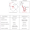
Figure 2. Properties and behaviors of stem cells that renew the alveoli and intestine
Stem cell features and renewal activity are compared between the alveolar and small intestinal epithelium. The schematic shows the bifunctional nature of the AT2 cell compared to the relatively ‘undifferentiated’ intestinal stem cell, and the table below contrasts other features of alveolar and intestinal stem cells. In the alveoli, AT2 stem cells (bright red) self-duplicate and transdifferentiate into flat AT1 cells (faint red) intermittently during aging. They also constitutively produce and secrete surfactant phospholipid into the alveolar airspace to reduce surface tension. In the intestine, there are two populations of intestinal stem cells (both shown as bright red). A major population regularly proliferates and generates ‘transit amplifying’ cells that flow upwards while continuing to proliferate and differentiate (faint red). The minor population is quiescent during renewal, serving as a back-up pool to regenerate primary stem cells that may be lost with injury.
Figure 3
Figure 3. Two models for the cellular mechanism of alveolar renewal and repair by AT2 cells
Rare AT2 cells (about 1% of the population) generate slowly expanding alveolar renewal foci during aging. Whether they are a molecularly distinct subset is unknown, but it has been shown that renewal foci are monoclonal, deriving from a single AT2 cell out of a pool of many. Two models are proposed to explain this monoclonal dominance. In one scenario (Model 1), a specialized AT2 stem cell intermittently proliferates to replace nearby dying cells (dashed circle) during aging. In the second scenario (Model 2), an AT2 cell is initially stochastically selected (thick green circle) then remains the dominant stem cell during aging by suppressing nearby AT2 cells (inhibitory arrow). In both models, acute injury with substantial alveolar cell loss results in activation of quiescent AT2 cells as ancillary progenitors (thick light green circles) to replace lost cells. Because they are quiescent during aging, their activation may involve either de novo initiation of an alveolar ‘injury’ signal that is not expressed during aging, downregulation of a suppressive signal, or a combination.
Figure 4
Figure 4. A hierarchy of cellular programs for local replacement of alveolar cells
A summary of the cellular mechanisms for renewing and repairing alveolar epithelium. (A) The cartoon indicates the relevant cell types depicted below. (B) Three distinct cellular programs for renewal and repair are schematized in order of deployment. Cells are represented as follows: solid grey fill, resting cells; dotted grey outlines, dead or dying cells; dark colors, proliferating cells; faint colors, progeny of proliferating cells. Top row: during aging, the sporadic loss of an alveolar cell (e.g. an AT1 cell shown here) triggers proliferation of an AT2 stem cell (red). In this example, a daughter cell transdifferentiates into an AT1 cell to replace the one that was lost. Middle row: if an injury locally depletes more alveolar cells than the AT2 stem cell can rapidly replace, ancillary AT2 cells (green) are induced to proliferate and transdifferentiate. Bottom row: if an injury locally depletes all AT2 cells, the LNEP/DASC population (blue) is activated. These cells extensively proliferate, migrate, and differentiate into Club and AT2 cells, which presumably generate other bronchiolar lineages and AT1 cells, respectively.
Figure 5
Figure 5. Lineage transitions between lung cell types observed during aging, after injury, and in cancer
A summary of reported in vivo lineage transitions between lung cell types in different contexts. Lineage transitions observed during renewal and following mild injury are classified as ‘constitutive’ (curved black arrows). Phenotypic switches only observed during repair or regeneration are classified as ‘facultative’ (curved colored arrows). Arrows are color-coded to indicate the specific cells targeted and experimental manipulations that elicited the indicated lineage conversion, described in text box. Straight black arrows indicate cell types of origin for specific cancer subtypes, and histologic and molecular transformations observed in these cancers. Grey filled boxes indicate cell populations that are self-maintaining during renewal. ‘?’ indicates uncertainty whether the indicated lineage switch occurs due to the inability to specifically mark and trace these cell types (see text for details).

Similar articles

Cited by

References

    1. Bertalanffy FD, Leblond CP. Structure of respiratory tissue. Lancet. 1955;269:1365–8. - PubMed
    1. Kauffman SL. Cell proliferation in the mammalian lung. Int Rev Exp Pathol. 1980;22:131–91. - PubMed
    1. Uhal BD. Cell cycle kinetics in the alveolar epithelium. Am J Physiol. 1997;272:L1031–45. - PubMed
    1. Leblond CP. Classification of Cell Populations on the Basis of Their Proliferative Behavior. Natl Cancer I Monogr. 1964;14:119–50. - PubMed
    1. Buckingham ME, Meilhac SM. Tracing cells for tracking cell lineage and clonal behavior. Dev Cell. 2011;21:394–409. - PubMed

Publication types

LinkOut - more resources