

Robot-aided in vitro measurement of patellar stability with consideration to the influence of muscle loading
- PMID: 26201401
- PMCID: PMC4511523
- DOI: 10.1186/s12938-015-0068-7
Robot-aided in vitro measurement of patellar stability with consideration to the influence of muscle loading
Abstract
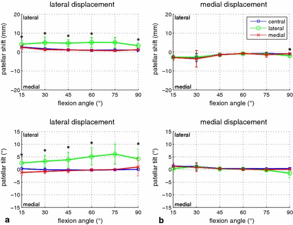
Background: Anterior knee pain is often associated with patellar maltracking and instability. However, objective measurement of patellar stability under clinical and experimental conditions is difficult, and muscular activity influences the results. In the present study, a new experimental setting for in vitro measurement of patellar stability was developed and the mediolateral force-displacement behavior of the native knee analyzed with special emphasis on patellar tilt and muscle loading.
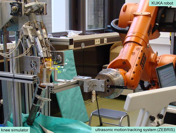
Methods: In the new experimental setup, two established testing methods were combined: an upright knee simulator for positioning and loading of the knee specimens, and an industry robot for mediolateral patellar displacement. A minimally invasive coupling and force control mechanism enabled unconstrained motion of the patella as well as measurement of patellar motion in all six degrees of freedom via an external ultrasonic motion-tracking system. Lateral and medial patellar displacement were measured on seven fresh-frozen human knee specimens in six flexion angles with varying muscle force levels, muscle force distributions, and displacement forces.
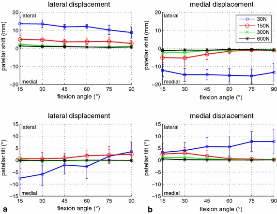
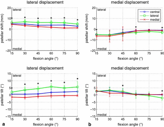
Results: Substantial repeatability was achieved for patellar shift (ICC(3,1) = 0.67) and tilt (ICC(3,1) = 0.75). Patellar lateral and medial shift decreased slightly with increasing flexion angle. Additional measurement of patellar tilt provided interesting insights into the different displacement mechanisms in lateral and medial directions. For lateral displacement, the patella tilted in the same (lateral) direction, and tilted in the opposite direction (again laterally) for medial displacement. With regard to asymmetric muscle loading, a significant influence (p < 0.03, up to 5 mm shift and 8° tilt) was found for lateral displacement and a reasonable relationship between muscle and patellar force, whereas no effect was visible in the medial direction.
Conclusion: The developed experimental setup delivered reproducible results and was found to be an excellent testing method for the in vitro analysis of patellar stability and future investigation of surgical techniques for patellar stabilization and total knee arthroplasty. We demonstrated a significant influence of asymmetric quadriceps loading on patellar stability. In particular, increased force application on the vastus lateralis muscle led to a clear increase of lateral patellar displacement.
Figures

Similar articles
-
Quantitative measurement of patellofemoral joint stability: force-displacement behavior of the human patella in vitro.J Orthop Res. 2003 Sep;21(5):780-6. doi: 10.1016/S0736-0266(03)00061-5. J Orthop Res. 2003. PMID: 12919863
-
The influence of asymmetric quadriceps loading on patellar tracking--an in vitro study.Knee. 2012 Dec;19(6):818-22. doi: 10.1016/j.knee.2012.04.011. Epub 2012 May 25. Knee. 2012. PMID: 22633902
-
Biomechanical effects of patellar positioning on intraoperative knee joint gap measurement in total knee arthroplasty.Clin Biomech (Bristol). 2010 May;25(4):352-8. doi: 10.1016/j.clinbiomech.2010.01.005. Epub 2010 Feb 1. Clin Biomech (Bristol). 2010. PMID: 20117864
-
Lateral force-displacement behaviour of the human patella and its variation with knee flexion--a biomechanical study in vitro.J Biomech. 1998 Dec;31(12):1147-52. doi: 10.1016/s0021-9290(98)00125-0. J Biomech. 1998. PMID: 9882047
-
Measurement of patellar tracking: assessment and analysis of the literature.Clin Orthop Relat Res. 2003 Jul;(412):241-59. doi: 10.1097/01.blo.0000068767.86536.9a. Clin Orthop Relat Res. 2003. PMID: 12838076 Review.
Cited by
-
Biomechanical evaluation of patellar tendon repair using Krackow suture technique.Biomed Eng Online. 2019 May 22;18(1):64. doi: 10.1186/s12938-019-0680-z. Biomed Eng Online. 2019. PMID: 31118104 Free PMC article.
-
Arthroscopic lateral retinacular release improves patello-femoral and femoro-tibial kinematics in patients with isolated lateral retinacular tightness.Knee Surg Sports Traumatol Arthrosc. 2022 Mar;30(3):791-799. doi: 10.1007/s00167-021-06434-w. Epub 2021 Jan 26. Knee Surg Sports Traumatol Arthrosc. 2022. PMID: 33496826 Free PMC article.
References
-
- Motsis EK, Paschos N, Pakos EE, Georgoulis AD. Review article: patellar instability after total knee arthroplasty. J Orthop Surg (HongKong). 2009;17(3):351–357. - PubMed
Publication types
MeSH terms
LinkOut - more resources
Full Text Sources
Other Literature Sources
