

How do stone attenuation and skin-to-stone distance in computed tomography influence the performance of shock wave lithotripsy in ureteral stone disease?
- PMID: 26201514
- PMCID: PMC4511972
- DOI: 10.1186/s12894-015-0069-7
How do stone attenuation and skin-to-stone distance in computed tomography influence the performance of shock wave lithotripsy in ureteral stone disease?
Abstract
Background: Shock wave lithotripsy (SWL) is a noninvasive, safe, and efficient treatment option for ureteral stones. Depending on stone location and size, the overall stone-free rate (SFR) varies significantly. Failure of stone disintegration results in unnecessary exposure to shock waves and radiation and requires alternative treatment procedures, which increases medical costs. It is therefore important to identify predictors of treatment success or failure in patients who are potential candidates for SWL before treatment. Nowadays, noncontrast computed tomography (NCCT) provides reliable information on stone location, size, number, and total stone burden. The impact of additional information provided by NCCT, such as skin-to-stone distance (SSD) and mean attenuation value (MAV), on stone fragmentation in ureteral stone disease has hardly been investigated separately so far. Thus, the objective of this study was to assess the influence of stone attenuation, SSD and body mass index (BMI) on the outcome of SWL in ureteral stones.
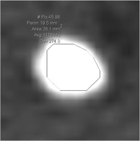
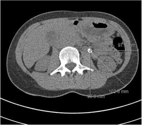
Methods: We reviewed the medical records of 104 patients (80 men, 24 women) with ureteral stone disease treated consecutively at our institution with SWL between 2010 and 2013. MAV in Hounsfield Units (HU) and SSD were determined by analyzing noncontrast computed tomography images. Outcome of SWL was defined as successful (visible stone fragmentation on kidney, ureter, and bladder film (KUB)) or failed (absent fragmentation on KUB).
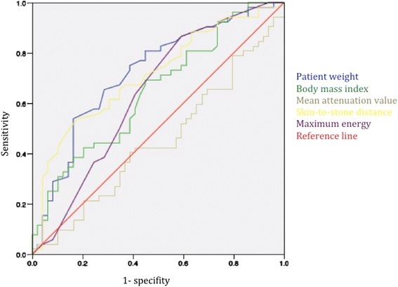
Results: Overall success of SWL was 50% (52 patients). Median stone attenuation was 956.9 HU (range 495-1210.8) in the group with successful disintegration and 944.6 (range 237-1302) in the patients who had absent or insufficient fragmentation. Median SSD was 125 mm (range 81-165 mm) in the group treated successfully and 141 mm (range 108-172 mm) in the patients with treatment failure. Unlike MAV (p = 0.37), SSD (p < 0.001) and BMI (p = 0.008) significantly correlated with treatment outcome.
Conclusion: The choice of treatment for ureteral stones should be based on stone location and size as considered in the AUA and EAU guidelines on urinary stone disease. In ambiguous cases, SSD and BMI can be used to assist in the decision. In this study, MAV showed no correlation with fragmentation rate of SWL.
Figures
References
-
- El-Nahas AR, El-Assmy AM, Mansour O, Sheir KZ. A prospective multivariate analysis of factors predicting stone disintegration by extracorporeal shock wave lithotripsy: the value of high-resolution noncontrast computed tomography. Eur Urol. 2007;51:1688. doi: 10.1016/j.eururo.2006.11.048. - DOI - PubMed
Publication types
MeSH terms
LinkOut - more resources
Full Text Sources
Other Literature Sources
Medical
