Randomized Controlled Trial
. 2015 Jul 23:16:310.
doi: 10.1186/s13063-015-0856-1.

Non-sedation versus sedation with a daily wake-up trial in critically ill patients receiving mechanical ventilation--effects on physical function: study protocol for a randomized controlled trial: a substudy of the NONSEDA trial

Affiliations
Randomized Controlled Trial

Non-sedation versus sedation with a daily wake-up trial in critically ill patients receiving mechanical ventilation--effects on physical function: study protocol for a randomized controlled trial: a substudy of the NONSEDA trial

Helene Korvenius Nedergaard et al. Trials. .

Abstract

Background: Critically ill patients rapidly loose much of their muscle mass and strength. This can be attributed to prolonged admission, prolonged mechanical ventilation and increased mortality, and it can have a negative impact on the degree of independence and quality of life. In the NONSEDA trial we randomize critically ill patients to non-sedation or sedation with a daily wake-up trial during mechanical ventilation in the intensive care unit. It has never been assessed whether non-sedation affects physical function. The aim of this study is to assess the effects of non-sedation versus sedation with a daily wake-up trial on physical function after discharge from intensive care unit.

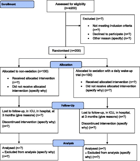
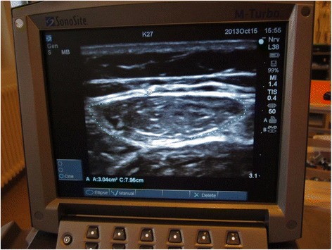
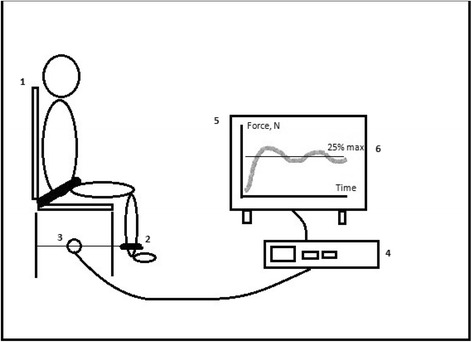
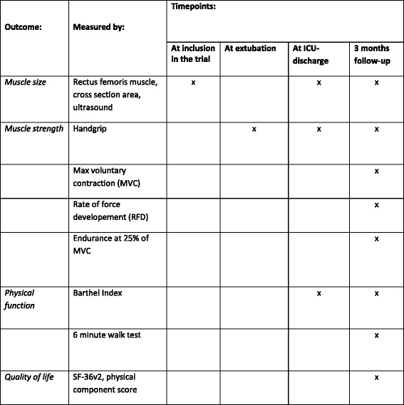
Methods/design: Investigator-initiated, randomized, clinical, parallel-group, superiority trial, including 700 patients in total, with a substudy concerning 200 of these patients. Inclusion criteria will be intubated, mechanically ventilated patients with expected duration of mechanical ventilation >24 h. Exclusion criteria will be patients with severe head trauma, coma at admission or status epilepticus, patients treated with therapeutic hypothermia, patients with PaO2/FiO2<9 where sedation might be necessary to ensure sufficient oxygenation or placing the patient in a prone position. The experimental intervention will be non-sedation supplemented with pain management during mechanical ventilation. The control intervention will be sedation with a daily wake-up trial. The co-primary outcome will be quality of life regarding physical function (SF-36, physical component) and degree of independence in activities of daily living (Barthel Index), and this will be assessed for all 700 patients participating in the NONSEDA trial. The secondary outcomes, which will be assessed for the subpopulation of 200 NONSEDA patients in the trial site, Kolding, will be 6-min walking distance, handgrip strength, muscle size (ultrasonographic measurement of the rectus femoris muscle cross-sectional area) and biomechanical data on lower extremity function (maximal voluntary contraction, rate of force development and endurance).

Discussion: This study is the first to investigate the effect of no sedation during critical illness on physical function. If an effect is found, it will add important information on how to prevent muscle weakness following critical illness.

Trial registration: The study has been approved by the relevant scientific ethics committee and is registered at ClinicalTrials.gov (ID: NCT02034942, 9 January 2014).


Figures

Fig. 1
Fig. 1
Flowchart. Schematic presentation of the patient flow through the trial
Fig. 2
Fig. 2
Ultrasonographic measurement of the rectus femoris muscle cross-sectional area. The rectus femoris muscle is part of the quadriceps muscle. Patients will be placed in supine position with their back raised to 45 degrees, with their legs in passive extension. The transducer will be placed over the rectus femoris muscle, perpendicular to the long axis of the right thigh, not depressing the dermal surface. Measurements will be made at 2/3 of the distance from the anterior superior iliac spine to the superior patellar border. This distance will be defined when the patient is placed as noted above, not with the patient standing up, since this changes the distance. For the scan, a linear transducer will be used, flat footprint, 5–8 MHz. The muscle is identified visually and an ultrasonographic picture is taken. Using the ultrasonographic software, the outer edge of the muscle is marked, and the cross-sectional area is calculated using planimetry
Fig. 3
Fig. 3
Study setup for biomechanical measurements of the lower limbs. 1: Custom-made chair, where the participant will be seated, back straight, with 90 degree flexion in the hip, 90 degree flexion in the knee, to ensure that the lower leg is in a vertical position. A safety strap will be fastened at the hip and hands held in the lap. 2: A strap will be fastened around the ankle, ensuring that the rigid transducer arm (3) is horizontal. 3: Strain-gauge transducer. 4: Computer with software for data collection. 5: Screen, where the participant will get visual feedback during endurance testing. A line will be marked on the screen (6), representing 25 % of the participants’ maximal voluntary contraction (MVC). Made with inspiration from J.B. Poulsen and colleagues [47]. The following measurements will be performed: the maximal voluntary contraction (MVC) and rate of force development (RFD). The participant is carefully instructed, using the same wording every time, to “stretch your leg as forcefully and as quickly as possible.” When maximal tension has been reached, it is maintained for 1–2 s and then released. The peak of the force-time curve is the MVC. The steepest slope of the curve is the RFD. Endurance: 25 % of the participants’ MVC will be calculated, and a line will be depicted on the screen. The participant will be instructed to exert a force sufficient to reach the line, not more, not less. The participant will maintain this force for as long as possible, though maximally 3 min. The participant will have constant visual feedback and standardized verbal encouragement, if needed
Fig. 4
Fig. 4
Outcomes and time points. The primary, secondary and exploratory outcomes and relevant time points

Similar articles

Cited by

References

    1. Danish Intensive Care Database, DID. [in Danish] Available at: https://www.sundhed.dk/content/cms/12/4712_aarsrapport-2011-did-290612.pdf. Accessed September 10, 2013.
    1. Granja C, Amaro A, Dias C, Costa-Pereira A. Outcome of ICU survivors: a comprehensive review. The role of patient-reported outcome studies. Acta Anaesthesiol Scand. 2012;56:1092–103. doi: 10.1111/j.1399-6576.2012.02686.x. - DOI - PubMed
    1. Parry SM, Berney S, Koopman R, Bryant A, El-Ansary D, Puthucheary Z, et al. Early rehabilitation in critical care (eRiCC): functional electrical stimulation with cycling protocol for a randomised controlled trial. BMJ Open. 2012;2 doi: 10.1136/bmjopen-2012-001891. - DOI - PMC - PubMed
    1. Gerovasili V, Stefanidis K, Vitzilaios K, Karatzanos E, Politis P, Koroneos A, et al. Electrical muscle stimulation preserves the muscle mass of critically ill patients: a randomized study. Crit Care. 2009;13:R161. doi: 10.1186/cc8123. - DOI - PMC - PubMed
    1. Schefold J, Bierbrauer J, Weber-Carstens S. Intensive care unit-acquired weakness (ICUAW) and muscle wasting in critically ill patients with severe sepsis and septic shock. J Cachex Sarcopenia Muscle. 2010;1:147–57. doi: 10.1007/s13539-010-0010-6. - DOI - PMC - PubMed

Publication types

MeSH terms

Substances

Associated data

LinkOut - more resources