

Path-programmable water droplet manipulations on an adhesion controlled superhydrophobic surface
- PMID: 26202206
- PMCID: PMC4511949
- DOI: 10.1038/srep12326
Path-programmable water droplet manipulations on an adhesion controlled superhydrophobic surface
Abstract
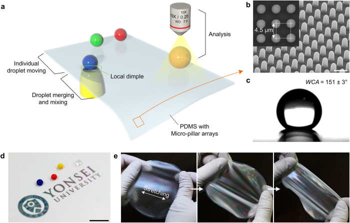
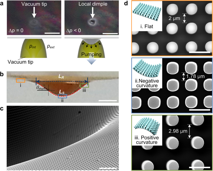
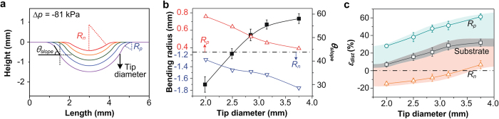
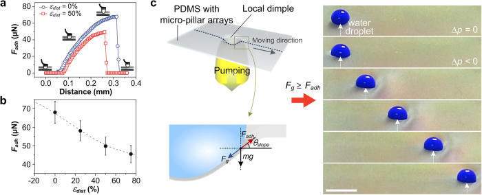
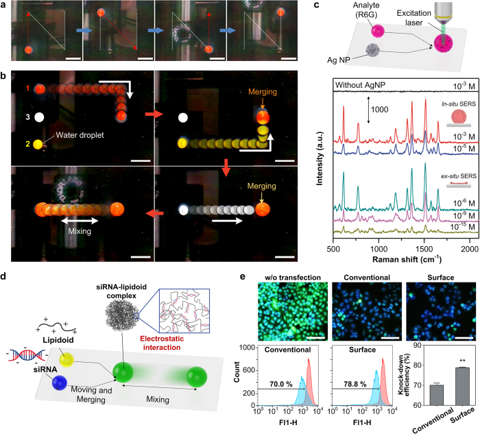
Here, we developed a novel and facile method to control the local water adhesion force of a thin and stretchable superhydrophobic polydimethylsiloxane (PDMS) substrate with micro-pillar arrays that allows the individual manipulation of droplet motions including moving, merging and mixing. When a vacuum pressure was applied below the PDMS substrate, a local dimple structure was formed and the water adhesion force of structure was significantly changed owing to the dynamically varied pillar density. With the help of the lowered water adhesion force and the slope angle of the formed dimple structure, the motion of individual water droplets could be precisely controlled, which facilitated the creation of a droplet-based microfluidic platform capable of a programmable manipulation of droplets. We showed that the platform could be used in newer and emerging microfluidic operations such as surface-enhanced Raman spectroscopy with extremely high sensing capability (10(-15) M) and in vitro small interfering RNA transfection with enhanced transfection efficiency of ~80%.
Figures
Similar articles
-
Droplet-driven transports on superhydrophobic-patterned surface microfluidics.Lab Chip. 2011 Nov 7;11(21):3642-8. doi: 10.1039/c1lc20390h. Epub 2011 Sep 14. Lab Chip. 2011. PMID: 21918770
-
Controllable adhesive superhydrophobic surfaces based on PDMS microwell arrays.Langmuir. 2013 Mar 12;29(10):3274-9. doi: 10.1021/la304492c. Epub 2013 Feb 25. Langmuir. 2013. PMID: 23391207
-
Splitting a droplet for femtoliter liquid patterns and single cell isolation.ACS Appl Mater Interfaces. 2015 May 6;7(17):9060-5. doi: 10.1021/am509177s. Epub 2015 Mar 29. ACS Appl Mater Interfaces. 2015. PMID: 25761507
-
Bioinspired super-antiwetting interfaces with special liquid-solid adhesion.Acc Chem Res. 2010 Mar 16;43(3):368-77. doi: 10.1021/ar900205g. Acc Chem Res. 2010. PMID: 19954162 Review.
-
Droplet based microfluidics.Rep Prog Phys. 2012 Jan;75(1):016601. doi: 10.1088/0034-4885/75/1/016601. Epub 2011 Dec 22. Rep Prog Phys. 2012. PMID: 22790308 Review.
Cited by
-
Micropillars in Cell Mechanobiology: Design, Fabrication, Characterization, and Biosensing Applications.Small Sci. 2024 Dec 9;5(4):2400410. doi: 10.1002/smsc.202400410. eCollection 2025 Apr. Small Sci. 2024. PMID: 40657190 Free PMC article. Review.
-
Superhydrophobic materials for biomedical applications.Biomaterials. 2016 Oct;104:87-103. doi: 10.1016/j.biomaterials.2016.06.050. Epub 2016 Jul 9. Biomaterials. 2016. PMID: 27449946 Free PMC article. Review.
-
Recent Developments in Artificial Super-Wettable Surfaces Based on Bioinspired Polymeric Materials for Biomedical Applications.Polymers (Basel). 2022 Jan 7;14(2):238. doi: 10.3390/polym14020238. Polymers (Basel). 2022. PMID: 35054645 Free PMC article. Review.
-
Mechanically Switchable Wetting Petal Effect in Self-Patterned Nanocolumnar Films on Poly(dimethylsiloxane).Nanomaterials (Basel). 2021 Sep 29;11(10):2566. doi: 10.3390/nano11102566. Nanomaterials (Basel). 2021. PMID: 34685004 Free PMC article.
-
Spatially and Temporally Controlled Hydrogels for Tissue Engineering.Mater Sci Eng R Rep. 2017 Sep;119:1-35. doi: 10.1016/j.mser.2017.07.001. Epub 2017 Jul 25. Mater Sci Eng R Rep. 2017. PMID: 29200661 Free PMC article.
References
-
- Daw R. & Finkelstein J. Lab on a chip. Nature 442, 367–367 (2006).
-
- deMello A. J. Control and detection of chemical reactions in microfluidic systems. Nature 442, 394–402 (2006). - PubMed
-
- El-Ali J., Sorger P. K. & Jensen K. F. Cells on chips. Nature 442, 403–411 (2006). - PubMed
-
- Yager P. et al. Microfluidic diagnostic technologies for global public health. Nature 442, 412–418 (2006). - PubMed
-
- Choi K., Ng A. H. C., Fobel R. & Wheeler A. R. Digital Microfluidics. Annu. Rev. Anal. Chem. 5, 413–440 (2012). - PubMed
Publication types
MeSH terms
Substances
LinkOut - more resources
Full Text Sources
Other Literature Sources
