. 2015 Oct;89(19):9896-908.
doi: 10.1128/JVI.01501-15. Epub 2015 Jul 22.

Brain Invasion by Mouse Hepatitis Virus Depends on Impairment of Tight Junctions and Beta Interferon Production in Brain Microvascular Endothelial Cells

Affiliations

Brain Invasion by Mouse Hepatitis Virus Depends on Impairment of Tight Junctions and Beta Interferon Production in Brain Microvascular Endothelial Cells

Christian Bleau et al. J Virol. 2015 Oct.

Abstract

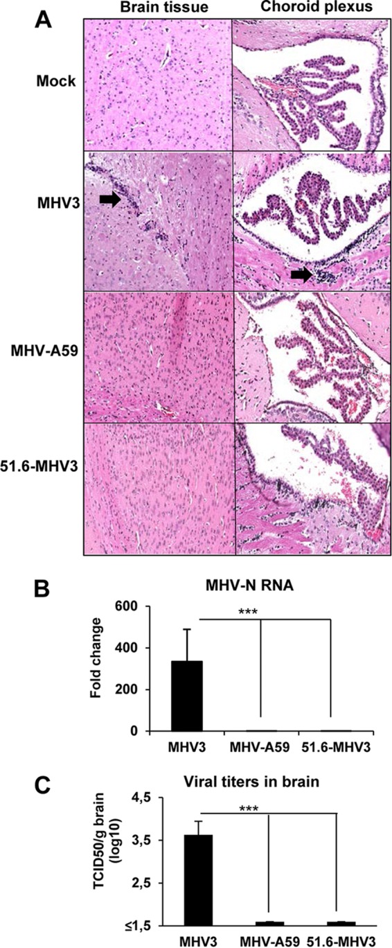
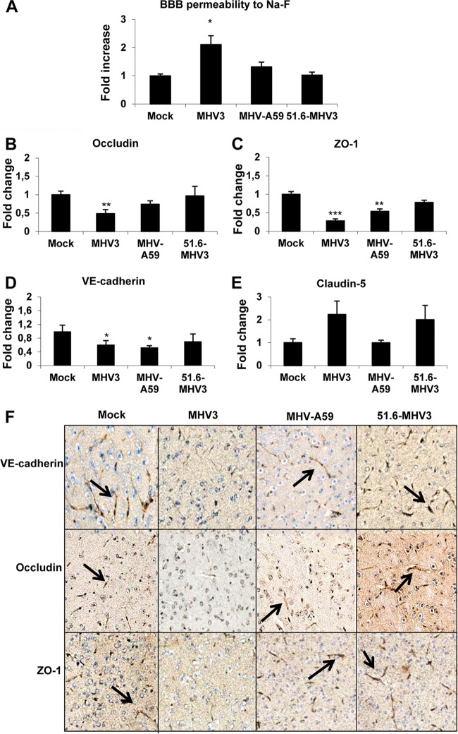
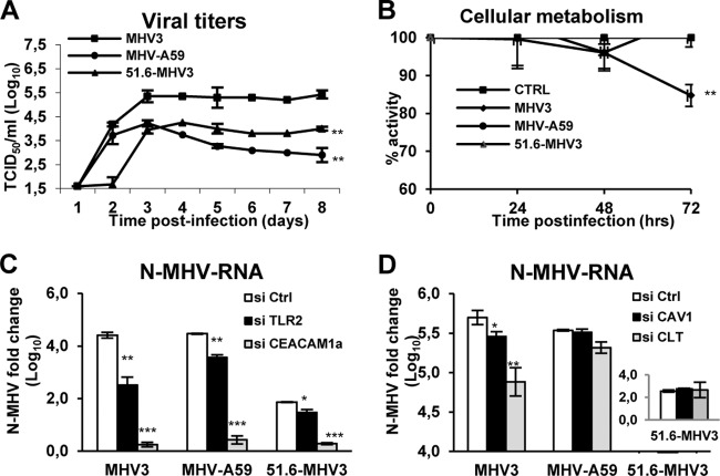
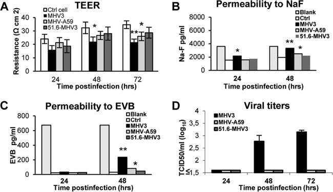
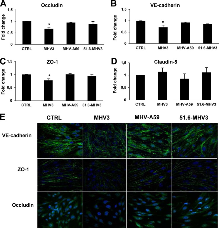
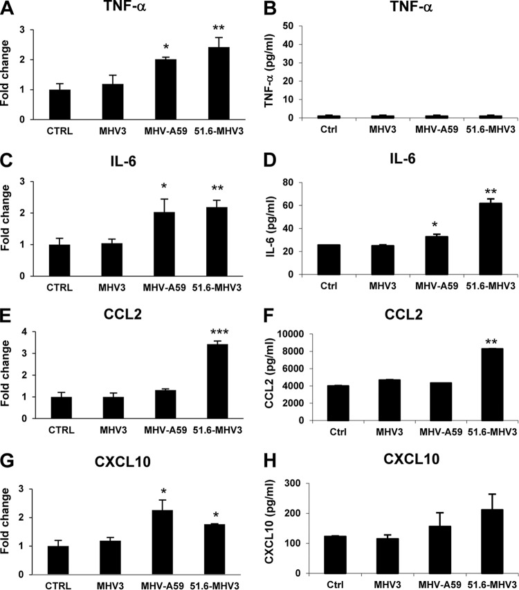
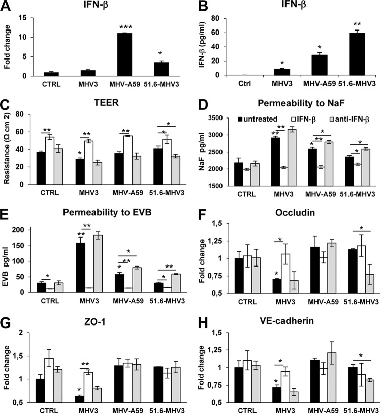
Coronaviruses (CoVs) have shown neuroinvasive properties in humans and animals secondary to replication in peripheral organs, but the mechanism of neuroinvasion is unknown. The major aim of our work was to evaluate the ability of CoVs to enter the central nervous system (CNS) through the blood-brain barrier (BBB). Using the highly hepatotropic mouse hepatitis virus type 3 (MHV3), its attenuated variant, 51.6-MHV3, which shows low tropism for endothelial cells, and the weakly hepatotropic MHV-A59 strain from the murine coronavirus group, we investigated the virus-induced dysfunctions of BBB in vivo and in brain microvascular endothelial cells (BMECs) in vitro. We report here a MHV strain-specific ability to cross the BBB during acute infection according to their virulence for liver. Brain invasion was observed only in MHV3-infected mice and correlated with enhanced BBB permeability associated with decreased expression of zona occludens protein 1 (ZO-1), VE-cadherin, and occludin, but not claudin-5, in the brain or in cultured BMECs. BBB breakdown in MHV3 infection was not related to production of barrier-dysregulating inflammatory cytokines or chemokines by infected BMECs but rather to a downregulation of barrier protective beta interferon (IFN-β) production. Our findings highlight the importance of IFN-β production by infected BMECs in preserving BBB function and preventing access of blood-borne infectious viruses to the brain.

Importance: Coronaviruses (CoVs) infect several mammals, including humans, and are associated with respiratory, gastrointestinal, and/or neurological diseases. There is some evidence that suggest that human respiratory CoVs may show neuroinvasive properties. Indeed, the severe acute respiratory syndrome coronavirus (SARS-CoV), causing severe acute respiratory syndrome, and the CoVs OC43 and 229E were found in the brains of SARS patients and multiple sclerosis patients, respectively. These findings suggest that hematogenously spread CoVs may gain access to the CNS at the BBB level. Herein we report for the first time that CoVs exhibit the ability to cross the BBB according to strain virulence. BBB invasion by CoVs correlates with virus-induced disruption of tight junctions on BMECs, leading to BBB dysfunction and enhanced permeability. We provide evidence that production of IFN-β by BMECs during CoV infection may prevent BBB breakdown and brain viral invasion.


Figures

FIG 1
FIG 1
Brain invasion by hematogenously spread MHVs correlates with virulence. (A) Groups of six or seven mice were i.p. mock infected (PBS) or infected with 1,000 TCID50 (50% tissue culture infective doses) of MHV3, MHV-A59, and 51.6-MHV3 for 72 h, and harvested brains were subjected to hematoxylin-eosin-safranin staining for histopathological analyses. (B) Viral detection of MHV3, MHV-A59, and 51.6-MHV3 at 72 h p.i. in brain samples of infected mice determined by qRT-PCR analysis of the viral nucleoprotein (N) expression and viral titration (TCID50). The viral detection threshold is 1.6 TCID50/ml. Values are means plus standard errors of means (error bars). Values that are significantly different (P < 0.001) are indicated (***).
FIG 2
FIG 2
MHV3, but not MHV-A59 and 51.6-MHV3, induces BBB breakdown. Groups of six or seven mice were i.p. mock infected (PBS) or infected with 1,000 TCID50 of MHV3, MHV-A59, and 51.6-MHV3 for 72 h. (A) Determination of BBB permeability assessed by sodium fluorescein (NaF) uptake 1 h prior to euthanasia. (B to E) mRNA expression of occludin (B), ZO-1 (C), VE-cadherin (D), and claudin-5 (E) was evaluated in brain samples by qRT-PCR. Values represent fold change in gene expression relative to mock-infected mice after normalization with HPRT expression. All samples were run in duplicate. Values that are significantly different from the value for mock-infected mice are indicated by asterisks as follows: *, P < 0.05; **, P < 0.01; ***, P < 0.001. (F) Immunohistochemistry stainings of VE-cadherin, occludin, and ZO-1 on brain sections of MHV3-, MHV-A59- and 51.6-MHV3-infected mice.
FIG 3
FIG 3
MHV3 exhibits higher tropism for BMECs than MHV-A59 and 51.6-MHV3 do. Mouse bEnd.3 immortalized brain microvascular endothelial cells (BMECs) were infected with MHV3, MHV-A59, or 51.6-MHV3 at an MOI of 0.1. (A) Kinetics of MHV infections up to 8 days p.i. (B) Metabolic activity of infected BMECs as determined by MTS-PMS colorimetric assay from 24 h to 72 h p.i. (C) Roles of CEACAM1a and TLR2 in MHV entry into BMECs. (D) Roles of caveolin-1 (CAV-1) and clathrin (CLT) in MHV endocytosis by BMECs. bEnd.3 cells were treated with specific siRNA to each molecule prior to infection for 24 h, and MHV nucleoprotein (N) mRNA expression was determined by qRT-PCR. All experiments were conducted in triplicate. Results are representative of two or three independent experiments. Values are means ± standard errors of means (error bars). Values that are significantly different are indicated by asterisks as follows: *, P < 0.05; **, P < 0.01; ***, P < 0.001. CTRL and Ctrl, control (uninfected cells).
FIG 4
FIG 4
MHV3, but not MHV-A59 and 51.6-MHV3, alters in vitro BMEC barrier integrity. An in vitro model of BBB was constructed using bEnd.3 cell monolayers grown on Transwell inserts, and cells were infected with MHV3, MHV-A59, or 51.6-MHV3 at an MOI of 0.1. (A) Transendothelial electrical resistance (TEER), reflecting barrier integrity, was monitored from 24 to 72 h. (B and C) Evaluation of the paracellular permeability of bEnd.3 monolayers to NaF (B) or Evans blue (C) dye at 24 and 48 h p.i. Blank corresponds to maximal permeability (insert without cells). (D) Determination of viral transmigration across the bEnd.3 monolayer. Viral titers were recorded in the basolateral chamber from 24 to 72 h p.i. The Viral detection threshold is 1.6 TCID50. All experiments were conducted in triplicate. Results are representative of two independent experiments. Values that are significantly different are indicated by asterisks as follows: *, P < 0.05; **, P < 0.01.
FIG 5
FIG 5
MHV3, but not MHV-A59 and 51.6-MHV3, alters tight-junction expression on BMECs. (A to D) bEnd.3 cells were infected for 48 h with MHV3, MHV-A59, and 51.6-MHV3 at an MOI of 0.1, and the levels of expression of occludin (A), VE-cadherin (B), ZO-1 (C), and claudin-5 (D) mRNA were evaluated by qRT-PCR. Values represent fold change in gene expression relative to uninfected cells (control [CTRL]) after normalization with HPRT expression. All samples were run in duplicate. All experiments were conducted in triplicate. Results are representative of two independent experiments. Values that are significantly different (P < 0.05) are indicated (*). (E) Immunofluorescence stainings of the tight-junction (green) VE-cadherin, occludin, and ZO-1 and the nucleus (blue) in uninfected (CTRL) and infected bEnd.3 cells. Images presented are from one representative experiment out of three independent experiments.
FIG 6
FIG 6
MHV3-induced breakdown of BMEC barrier does not depend on inflammatory factors. (A to H) bEnd.3 cells were infected with MHV3, MHV-A59, and 51.6-MHV3 at an MOI of 0.1, and mRNA expression (A, C, E, and G) and protein levels (B, D, F, and H) of TNF-α, IL-6, CCL2, and CXCL10 were evaluated by qRT-PCR and ELISAs, respectively, at 24 h p.i. All samples were run in duplicate. All experiments were conducted in triplicate. Results are representative of two independent experiments. Values that are significantly different are indicated by asterisks as follows: *, P < 0.05; **, P < 0.01; ***, P < 0.001.
FIG 7
FIG 7
MHV3-induced breakdown of BMEC barrier is related to downregulation of barrier protective IFN-β production by infected BMECs. (A and B) bEnd.3 cells were infected with MHV3, MHV-A59, and 51.6-MHV3 at an MOI of 0.1, and mRNA expression (A) and protein expression (B) of IFN-β was evaluated at 24 h p.i. by qRT-PCR and ELISAs, respectively. All samples were run in duplicate. All experiments were conducted in triplicate. (C to H) Evaluation of barrier integrity of bEnd.3 monolayers infected for 48 h with MHV3, MHV-A59, and 51.6-MHV3 at an MOI of 0.1 in the presence of recombinant IFN-β (100 pg/ml) or anti-IFN-β monoclonal antibodies (1 μg/ml). (C) Recordings of TEER in infected bEnd.3 cultures. (D and E) Evaluation of the paracellular permeability of infected bEnd.3 monolayers to NaF (D) or Evans blue (E) dyes. (F to H) mRNA expression levels of occludin (F), ZO-1 (G), and VE-cadherin (H) in bEnd.3 cells evaluated by qRT-PCR. Values represent fold change in gene expression relative to uninfected cells (control [CTRL]) after normalization with HPRT expression. Results are representative of two independent experiments. Values that are significantly different are indicated by asterisks as follows: *, P < 0.05; **, P < 0.01; ***, P < 0.001.

Similar articles

Cited by

References

    1. Spindler KR, Hsu TH. 2012. Viral disruption of the blood-brain barrier. Trends Microbiol 20:282–290. doi:10.1016/j.tim.2012.03.009. - DOI - PMC - PubMed
    1. Hase T, Dubois DR, Summers PL. 1990. Comparative study of mouse brains infected with Japanese encephalitis virus by intracerebral or intraperitoneal inoculation. Int J Exp Pathol 71:857–869. - PMC - PubMed
    1. Kobiler D, Lustig S, Gozes Y, Ben-Nathan D, Akov Y. 1989. Sodium dodecylsulphate induces a breach in the blood-brain-barrier and enables a West Nile virus variant to penetrate into mouse brain. Brain Res 496:314–316. doi:10.1016/0006-8993(89)91079-2. - DOI - PubMed
    1. Lustig S, Danenberg HD, Kafri Y, Kobiler D, Ben-Nathan D. 1992. Viral neuroinvasion and encephalitis induced by lipopolysaccharide and its mediators. J Exp Med 176:707–712. doi:10.1084/jem.176.3.707. - DOI - PMC - PubMed
    1. Wang T, Town T, Alexopoulou L, Anderson JF, Fikrig E, Flavell RA. 2004. Toll-like receptor 3 mediates West Nile virus entry into the brain causing lethal encephalitis. Nat Med 10:1366–1373. doi:10.1038/nm1140. - DOI - PubMed

Publication types

MeSH terms

LinkOut - more resources