Temporal Characterization of Marburg Virus Angola Infection following Aerosol Challenge in Rhesus Macaques
- PMID: 26202230
- PMCID: PMC4577920
- DOI: 10.1128/JVI.01147-15
Temporal Characterization of Marburg Virus Angola Infection following Aerosol Challenge in Rhesus Macaques
Abstract
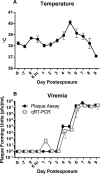
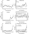
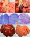
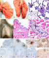
Marburg virus (MARV) infection is a lethal hemorrhagic fever for which no licensed vaccines or therapeutics are available. Development of appropriate medical countermeasures requires a thorough understanding of the interaction between the host and the pathogen and the resulting disease course. In this study, 15 rhesus macaques were sequentially sacrificed following aerosol exposure to the MARV variant Angola, with longitudinal changes in physiology, immunology, and histopathology used to assess disease progression. Immunohistochemical evidence of infection and resulting histopathological changes were identified as early as day 3 postexposure (p.e.). The appearance of fever in infected animals coincided with the detection of serum viremia and plasma viral genomes on day 4 p.e. High (>10(7) PFU/ml) viral loads were detected in all major organs (lung, liver, spleen, kidney, brain, etc.) beginning day 6 p.e. Clinical pathology findings included coagulopathy, leukocytosis, and profound liver destruction as indicated by elevated liver transaminases, azotemia, and hypoalbuminemia. Altered cytokine expression in response to infection included early increases in Th2 cytokines such as interleukin 10 (IL-10) and IL-5 and late-stage increases in Th1 cytokines such as IL-2, IL-15, and granulocyte-macrophage colony-stimulating factor (GM-CSF). This study provides a longitudinal examination of clinical disease of aerosol MARV Angola infection in the rhesus macaque model.
Importance: In this study, we carefully analyzed the timeline of Marburg virus infection in nonhuman primates in order to provide a well-characterized model of disease progression following aerosol exposure.
Copyright © 2015, American Society for Microbiology. All Rights Reserved.
Figures


Similar articles
-
Transcriptional Profiling of the Immune Response to Marburg Virus Infection.J Virol. 2015 Oct;89(19):9865-74. doi: 10.1128/JVI.01142-15. Epub 2015 Jul 22. J Virol. 2015. PMID: 26202234 Free PMC article.
-
Aerosol exposure to the angola strain of marburg virus causes lethal viral hemorrhagic Fever in cynomolgus macaques.Vet Pathol. 2010 Sep;47(5):831-51. doi: 10.1177/0300985810378597. Vet Pathol. 2010. PMID: 20807825
-
Virus-Like Particle Vaccination Protects Nonhuman Primates from Lethal Aerosol Exposure with Marburgvirus (VLP Vaccination Protects Macaques against Aerosol Challenges).Viruses. 2016 Apr 8;8(4):94. doi: 10.3390/v8040094. Viruses. 2016. PMID: 27070636 Free PMC article.
-
Marburg virus pathogenesis - differences and similarities in humans and animal models.Virol J. 2019 Dec 30;16(1):165. doi: 10.1186/s12985-019-1272-z. Virol J. 2019. PMID: 31888676 Free PMC article. Review.
-
Development of vaccines for Marburg hemorrhagic fever.Expert Rev Vaccines. 2007 Feb;6(1):57-74. doi: 10.1586/14760584.6.1.57. Expert Rev Vaccines. 2007. PMID: 17280479 Review.
Cited by
-
Dimerization Controls Marburg Virus VP24-dependent Modulation of Host Antioxidative Stress Responses.J Mol Biol. 2016 Aug 28;428(17):3483-94. doi: 10.1016/j.jmb.2016.07.020. Epub 2016 Aug 4. J Mol Biol. 2016. PMID: 27497688 Free PMC article.
-
Immune correlates of postexposure vaccine protection against Marburg virus.Sci Rep. 2020 Feb 20;10(1):3071. doi: 10.1038/s41598-020-59976-3. Sci Rep. 2020. PMID: 32080323 Free PMC article.
-
Post-exposure treatments for Ebola and Marburg virus infections.Nat Rev Drug Discov. 2018 Jun;17(6):413-434. doi: 10.1038/nrd.2017.251. Epub 2018 Jan 29. Nat Rev Drug Discov. 2018. PMID: 29375139 Review.
-
Filovirus Strategies to Escape Antiviral Responses.Curr Top Microbiol Immunol. 2017;411:293-322. doi: 10.1007/82_2017_13. Curr Top Microbiol Immunol. 2017. PMID: 28685291 Free PMC article. Review.
-
Emerging and reemerging infectious diseases: global trends and new strategies for their prevention and control.Signal Transduct Target Ther. 2024 Sep 11;9(1):223. doi: 10.1038/s41392-024-01917-x. Signal Transduct Target Ther. 2024. PMID: 39256346 Free PMC article. Review.
References
-
- Shu HL, Siegert R, Slenczka W. 1969. The pathogenesis and epidemiology of the “Marburg-virus” infection. Ger Med Monthly 14:7–10. - PubMed
Publication types
MeSH terms
Substances
Grants and funding
LinkOut - more resources
Full Text Sources
Other Literature Sources

