. 2015 Oct;89(19):10023-30.
doi: 10.1128/JVI.01530-15. Epub 2015 Jul 22.

The Cellular Factor NXP2/MORC3 Is a Positive Regulator of Influenza Virus Multiplication

Affiliations

The Cellular Factor NXP2/MORC3 Is a Positive Regulator of Influenza Virus Multiplication

Lorena S Ver et al. J Virol. 2015 Oct.

Erratum in

Abstract

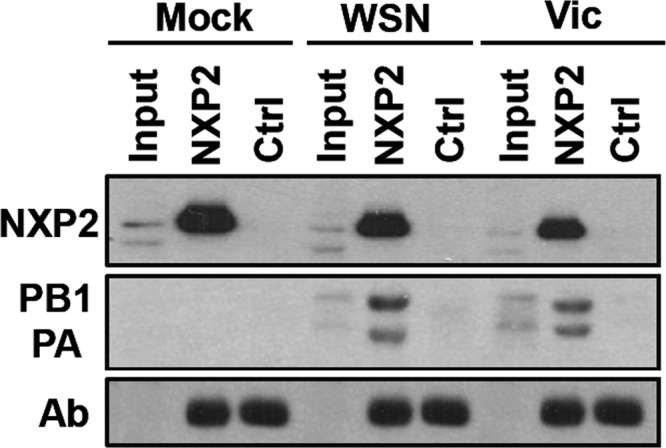
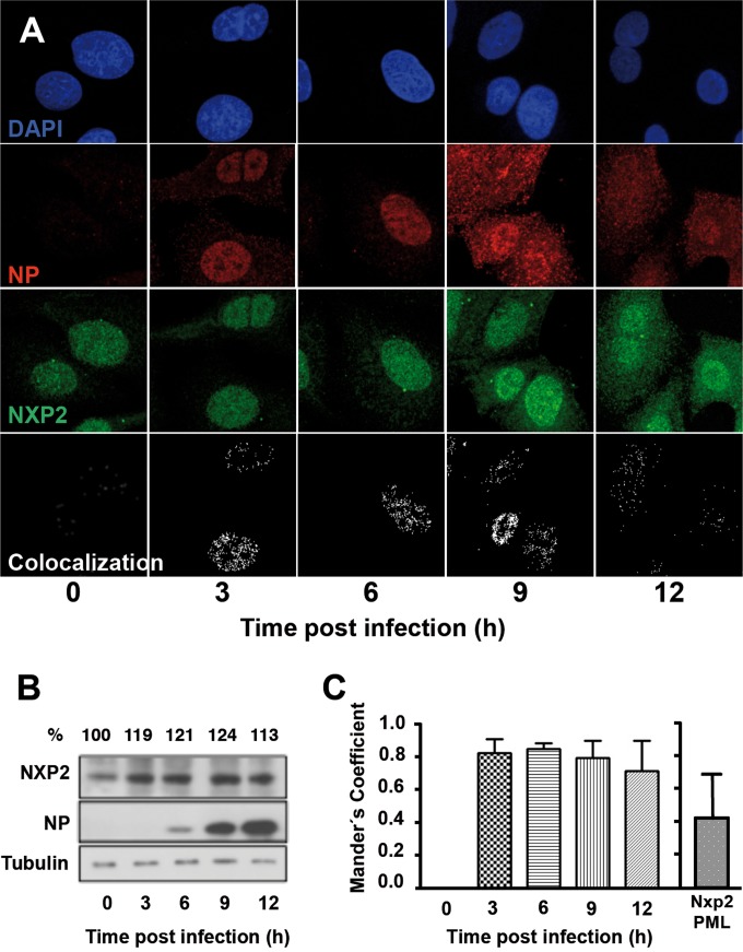
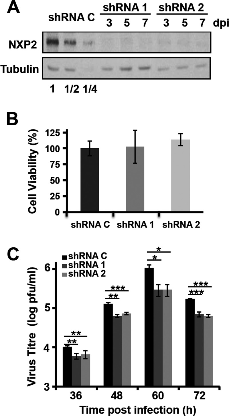
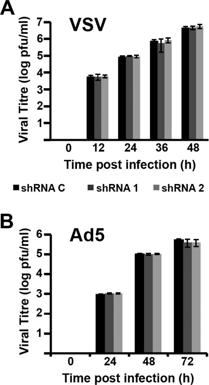
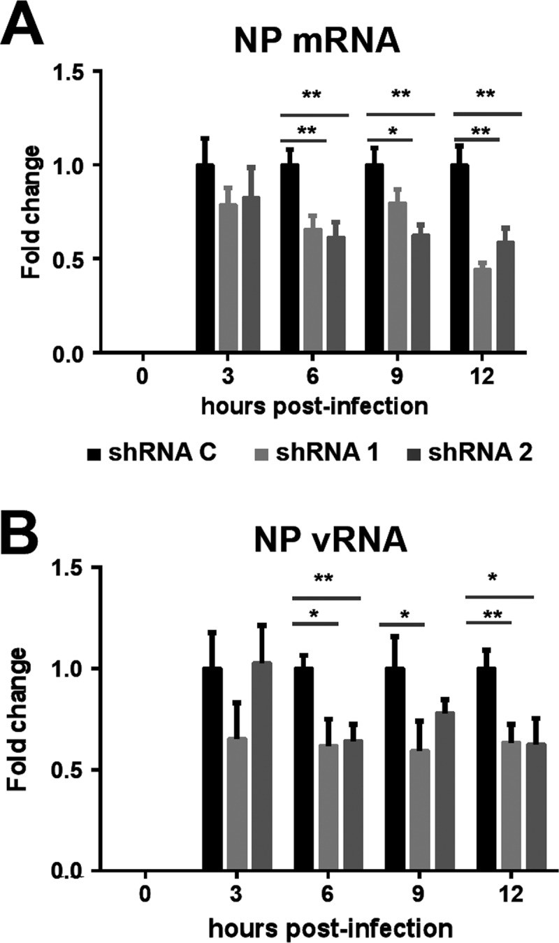
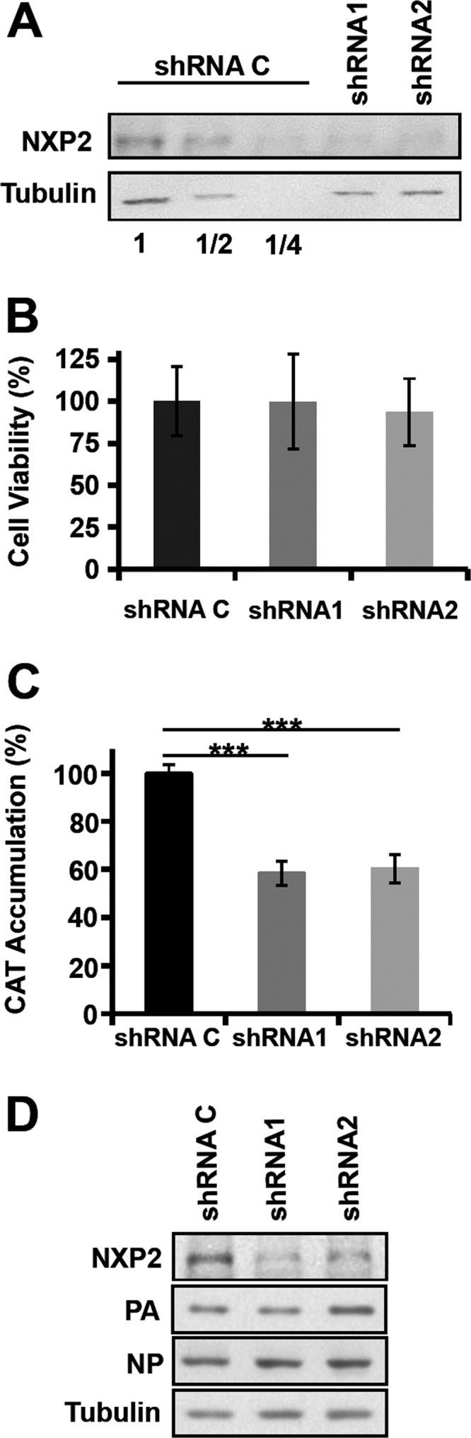
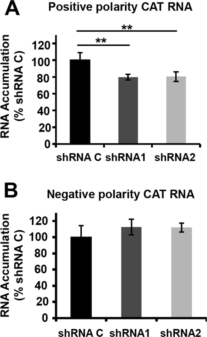
Transcription and replication of influenza A virus are carried out in the nuclei of infected cells in the context of viral ribonucleoproteins (RNPs). The viral polymerase responsible for these processes is a protein complex composed of the PB1, PB2, and PA proteins. We previously identified a set of polymerase-associated cellular proteins by proteomic analysis of polymerase-containing intracellular complexes expressed and purified from human cells. Here we characterize the role of NXP2/MORC3 in the infection cycle. NXP2/MORC3 is a member of the Microrchidia (MORC) family that is associated with the nuclear matrix and has RNA-binding activity. Influenza virus infection led to a slight increase in NXP2/MORC3 expression and its partial relocalization to the cytoplasm. Coimmunoprecipitation and immunofluorescence experiments indicated an association of NXP2/MORC3 with the viral polymerase and RNPs during infection. Downregulation of NXP2/MORC3 by use of two independent short hairpin RNAs (shRNAs) reduced virus titers in low-multiplicity infections. Consistent with these findings, analysis of virus-specific RNA in high-multiplicity infections indicated a reduction of viral RNA (vRNA) and mRNA after NXP2/MORC3 downregulation. Silencing of NXP2/MORC3 in a recombinant minireplicon system in which virus transcription and replication are uncoupled showed reductions in cat mRNA and chloramphenicol acetyltransferase (CAT) protein accumulation but no alterations in cat vRNA levels, suggesting that NXP2/MORC3 is important for influenza virus transcription.

Importance: Influenza virus infections appear as yearly epidemics and occasional pandemics of respiratory disease, with high morbidity and occasional mortality. Influenza viruses are intracellular parasites that replicate and transcribe their genomic ribonucleoproteins in the nuclei of infected cells, in a complex interplay with host cell factors. Here we characterized the role of the human NXP2/MORC3 protein, a member of the Microrchidia family that is associated with the nuclear matrix, during virus infection. NXP2/MORC3 associates with the viral ribonucleoproteins in infected cells. Downregulation of NXP2/MORC3 reduced virus titers and accumulations of viral genomic RNA and mRNAs. Silencing of NXP2/MORC3 in an influenza virus CAT minireplicon system diminished CAT protein and cat mRNA levels but not genomic RNA levels. We propose that NXP2/MORC3 plays a role in influenza virus transcription.


Figures

FIG 1
FIG 1
Interaction of NXP2/MORC3 with influenza virus polymerase. Cultures of HEK293T cells were infected with the WSN or Victoria (Vic) influenza virus strain at an MOI of 3 PFU/cell or mock infected (Mock). At 6 hpi, total cell extracts were prepared and immunoprecipitated with anti-NXP2/MORC3 or control antibodies. The presence of NXP2/MORC3, PB1, and PA was examined by Western blotting with an antibody specific for NXP2/MORC3 or a mixture of anti-PB1 and anti-PA antibodies. Input, total cell extracts; NXP2, immunoprecipitates obtained with NXP2/MORC3-specific antibodies; Ctrl, immunoprecipitates obtained with unspecific antibodies. The positions of NXP2/MORC3, PB1, PA, and the IgG heavy chain (Ab) are indicated to the left. Changes in electrophoretic mobility for the PB1 and PA proteins are a consequence of the immunoprecipitation process.
FIG 2
FIG 2
Accumulation and intracellular localization of NXP2/MORC3 during influenza virus infection. (A) Cultures of A549 cells were infected with the WSN influenza virus at an MOI of 3 PFU/cell. At the indicated times after infection, cells were fixed and processed for confocal immunofluorescence microscopy. Representative optical sections from three independent experiments are shown. Nuclei are presented in blue, NP in red, and NXP2/MORC3 in green. Colocalization masks are shown in white. (B) Total cell extracts of parallel cultures were analyzed by Western blotting with antibodies specific for NXP2/MORC3, NP, and β-tubulin (loading marker). Representative data from one of three independent experiments are presented. Numbers at the top indicate the quantification of the NXP2/MORC3 signal, using the value obtained at the time of virus inoculation as 100%. (C) Quantification of NP-NXP2/MORC3 colocalization. ImageJ software was used to calculate Manders's coefficient for colocalization. Averages for the values from a representative experiment are shown. Error bars indicate standard deviations. As a specificity control, the panel on the right shows Manders's coefficient for colocalization of NXP2 with PML, as an example of an alternative nuclear protein.
FIG 3
FIG 3
Kinetics of influenza virus multiplication in NXP2/MORC3-downregulated cells. Cultures of A549 cells were transduced with lentiviral vectors expressing an irrelevant short hairpin RNA (shRNA C) or shRNAs targeting the NXP2/MORC3 mRNA (shRNA1 or shRNA2). When the accumulation levels of NXP2/MORC3 were reduced, the cultures were infected with influenza virus WSN at an MOI of 0.001 PFU/cell. (A) Western blot analysis of NXP2/MORC3 at the indicated days post-lentiviral inoculation (dpi), using β-tubulin as a loading control. Serial 2-fold dilutions of control extracts were loaded for improved quantification. (B) The viability of control or downregulated A549 cells was determined using an MTT-based colorimetric assay as described in Materials and Methods. (C) The influenza virus infectivity of the supernatant was determined by plaque assay at the indicated times postinfection. The results presented are representative of three independent experiments. The statistical significance of the differences from the control data set was determined using the Student t test (*, P < 0.05; **, P < 0.01; ***, P < 0.001).
FIG 4
FIG 4
Kinetics of VSV and adenovirus multiplication in NXP2/MORC3-downregulated cells. Cultures of A549 cells were transduced with lentiviral vectors expressing an irrelevant short hairpin RNA (shRNA C) or shRNAs targeting NXP2/MORC3 mRNA (shRNA1 or shRNA2). When the accumulation levels of NXP2/MORC3 were reduced, the cultures were infected with VSV (top; MOI = 0.001 PFU/cell) or Ad5 (bottom; MOI = 5 PFU/cell) as described in Materials and Methods. Virus titration was performed with samples obtained at the indicated times. The results presented are representative of two or three independent experiments.
FIG 5
FIG 5
Accumulation of viral genomic RNA and mRNAs during infection of NXP2/MORC3-downregulated cells. Cultures of A549 cells were transduced with lentiviral vectors expressing an irrelevant short hairpin RNA (shRNA C) or shRNAs targeting NXP2/MORC3 mRNA (shRNA1 or shRNA2). When the accumulation levels of NXP2/MORC3 were reduced, the cultures were infected with influenza virus WSN at an MOI of 3 PFU/cell. Total cell RNA was isolated at the indicated times after infection, and the accumulation of NP mRNA (A) and vRNA (B) was determined by RT-qPCR, using the conditions and primers described by Kawakami et al. (45). The values shown are averages and standard deviations for three determinations and have been standardized to the value obtained for control cells at each infection time point. The statistical significance of differences from the data obtained for control cells was determined using the Student t test (*, P < 0.05; **, P < 0.01).
FIG 6
FIG 6
Biological activity of recombinant influenza virus RNPs in NXP2/MORC3-downregulated cells. (A) Cultures of HEK293T cells were transduced with lentiviral vectors expressing an irrelevant short hairpin RNA (shRNA C) or shRNAs targeting NXP2/MORC3 mRNA (shRNA1 or shRNA2), and silencing was monitored in total cell extracts by Western blotting. Serial 2-fold dilutions of control extracts were loaded for improved quantification. The accumulation of β-tubulin was used as a loading control. (B) The viability of control or NXP2/MORC3-downregulated HEK293T cells was tested by using an MTT-based colorimetric assay as described in Materials and Methods. (C) The NXP2/MORC3-downregulated or control HEK293T cells described for panel A were used for in vivo RNP reconstitution. The cultures were transfected with plasmids expressing the polymerase subunits and the NP, as well as with a genomic plasmid expressing a pseudoviral genome containing the cat gene in negative polarity. Twenty-four hours after transfection, the accumulation of the CAT protein in total cell extracts was analyzed by ELISA. The statistical significance of differences from the control data set was determined using the Student t test (***, P < 0.001). (D) Aliquots of the samples used in the experiment whose results are shown in panel C were also analyzed for the presence of RNPs by Western blotting, using β-tubulin as a loading control.
FIG 7
FIG 7
Transcription and replication activity of recombinant influenza virus RNPs in NXP2/MORC3-downregulated cells. Cultures of HEK293T cells were transduced with lentiviral vectors expressing an irrelevant short hairpin RNA (shRNA C) or shRNAs targeting NXP2/MORC3 mRNA (shRNA1 or shRNA2). When the expression of NXP2/MORC3 was downregulated, the cultures were transfected with plasmids expressing the polymerase subunits and the NP, as well as with a genomic plasmid expressing a pseudoviral genome containing the cat gene in negative polarity. At 24 h posttransfection, total cell RNA was isolated. (A) The accumulation of positive-polarity RNA was determined by hybridization with CAT-specific probes and is presented as a percentage of the maximal value obtained for shRNA C-silenced cells. (B) The accumulation of negative-polarity RNA was determined by hybridization with CAT-specific probes and is presented as a percentage of the maximal value obtained for shRNA C-silenced cells. The values represent the averages and standard deviations for three determinations. The statistical significance was determined by the Student t test (**, P < 0.01).

Similar articles

Cited by

References

    1. Palese P, Shaw M. 2013. Orthomyxoviridae, 6th ed, vol 1 Lippincott Williams & Wilkins, Philadelphia, PA.
    1. Vasin AV, Temkina OA, Egorov VV, Klotchenko SA, Plotnikova MA, Kiselev OI. 2014. Molecular mechanisms enhancing the proteome of influenza A viruses: an overview of recently discovered proteins. Virus Res 185:53–63. doi:10.1016/j.virusres.2014.03.015. - DOI - PubMed
    1. Arranz R, Coloma R, Chichon FJ, Conesa JJ, Carrascosa JL, Valpuesta JM, Ortin J, Martin-Benito J. 2012. The structure of native influenza virion ribonucleoproteins. Science 338:1634–1637. doi:10.1126/science.1228172. - DOI - PubMed
    1. Martin-Benito J, Ortin J. 2013. Influenza virus transcription and replication. Adv Virus Res 87:113–137. doi:10.1016/B978-0-12-407698-3.00004-1. - DOI - PubMed
    1. Resa-Infante P, Jorba N, Coloma R, Ortin J. 2011. The influenza virus RNA synthesis machine: advances in its structure and function. RNA Biol 8:207–215. doi:10.4161/rna.8.2.14513. - DOI - PMC - PubMed

Publication types

MeSH terms

LinkOut - more resources