. 2015 Jul 23:5:12465.
doi: 10.1038/srep12465.

Blockade of Wnt/β-catenin signaling suppresses breast cancer metastasis by inhibiting CSC-like phenotype

Affiliations

Blockade of Wnt/β-catenin signaling suppresses breast cancer metastasis by inhibiting CSC-like phenotype

Gyu-Beom Jang et al. Sci Rep. .

Abstract

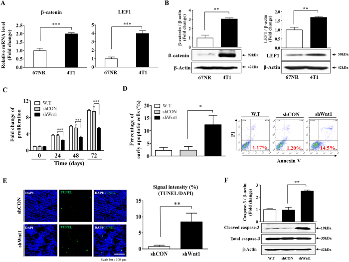
The identification of cancer stem cells (CSCs) represents an important milestone in the understanding of chemodrug resistance and cancer recurrence. More specifically, some studies have suggested that potential metastasis-initiating cells (MICs) might be present within small CSC populations. The targeting and eradication of these cells represents a potential strategy for significantly improving clinical outcomes. A number of studies have suggested that dysregulation of Wnt/β-catenin signaling occurs in human breast cancer. Consistent with these findings, our previous data have shown that the relative level of Wnt/β-catenin signaling activity in breast cancer stem cells (BCSCs) is significantly higher than that in bulk cancer cells. These results suggest that BCSCs could be sensitive to therapeutic approaches targeting Wnt/β-catenin signaling pathway. In this context, abnormal Wnt/β-catenin signaling activity may be an important clinical feature of breast cancer and a predictor of poor survival. We therefore hypothesized that Wnt/β-catenin signaling might regulate self-renewal and CSC migration, thereby enabling metastasis and systemic tumor dissemination in breast cancer. Here, we investigated the effects of inhibiting Wnt/β-catenin signaling on cancer cell migratory potential by examining the expression of CSC-related genes, and we examined how this pathway links metastatic potential with tumor formation in vitro and in vivo.


Conflict of interest statement

The authors declare no competing financial interests.

Figures

Figure 1
Figure 1. Comparison of Wnt/β-catenin signaling-related genes in malignant breast cancer tissues and their normal counterparts.
Cancerous and non-cancerous breast tissues (kindly provided by Dr. Lee at the National Cancer Center, Korea) were stained with antibodies against Wnt1. Wnt1 was expressed to a greater extent in the cancerous tissues than in the non-cancerous tissues. DAPI staining was performed to label the nuclei within each field (A). A significant correlation between poor prognosis and the expression of a negative (GSK3β) or positive (TCF4) regulator of Wnt/β-catenin signaling was observed in the human breast cancer datasets assessed, which were obtained through the Oncomine dataset repository (www.oncomine.org) (B). The results are presented as the mean ± SD, as determined from more than three independent experiments. *P < 0.05, **P < 0.01, ***P < 0.001.
Figure 2
Figure 2. Constitutive activation of the Wnt/β-catenin signaling pathway is a hallmark of tumorigenicity and maintenance of BCSCs.
67NR cells form primary tumors readily, although the tumor cells do not intravasate. On the other hand, 4T1 cells have full metastatic properties (A). The percentages of LEF1, cyclin D1, TCF-4, and β-catenin-positive cells in both Aldefluor-positive (B) and Sca-1-positive (C) subpopulations of non-invasive 67NR cells and highly invasive 4T1 cells were evaluated by flow cytometric analysis (B,C). Wnt3a-induced Wnt/β-catenin signaling in ALDH1-positive BCSC subpopulations was assessed using a TOP Flash luciferase reporter. Wnt3a treatment induced transcriptional activity to a greater extent in the ALDH1-positive BCSC subpopulations compared with that in the ALDH1-negative subpopulations (D). Wnt1 knockdown inhibited the tumor sphere formation of 4T1 cells. Spheres that were greater than 100 μm in size were enumerated, and a representative image of a tumor sphere is shown. The averages of three independent experiments are shown (E). Wnt1 knockdown led to a decrease in the percentage of CD44+/CD24 cells as a proportion of the total cancer cells (F). Abbreviations: TSFE, tumor sphere-forming efficiency. The results are presented as the mean ± SD, as determined from three independent experiments. *P < 0.05, **P < 0.01, ***P < 0.001.
Figure 3
Figure 3. Down-regulation of Wnt/β-catenin signaling suppresses tumor growth.
The relative expression of LEF1 and β-catenin, which are downstream components of Wnt/β-catenin signaling, in both non-invasive 67NR cells and highly invasive 4T1 cells was evaluated by real-time PCR and western blotting (A,B). Transfection of cells with Wnt1 shRNA led to a time-dependent decrease in the number of cells compared with that observed following transfection with control shRNA (C). Wnt1 knockdown-mediated cytotoxicity was evaluated by flow cytometry using PE-labeled annexin-V (D). Wnt1 knockdown-mediated apoptotic DNA fragmentation and condensation were visualized by TUNEL assay (E). The level of activated (cleaved) caspase-3 in cells undergoing Wnt1 knockdown-induced apoptosis was evaluated by western blot, using an antibody targeted against activated caspase-3 (F). DAPI staining was performed to label the nuclei within each field. β-actin was used as an internal control. The results are presented as the mean ± SD, as determined from three independent experiments. *P < 0.05, **P < 0.01, ***P < 0.001.
Figure 4
Figure 4. The Wnt/β-catenin signaling pathway regulates tumor cell invasion.
Cell migration ability was evaluated by transwell migration assay. Transfection with Wnt1 shRNA significantly decreased 4T1 cell migration across the membrane in both the upper and lower compartments of transwells compared with that observed following transfection with control shRNA (A,B). Wnt1 knockdown-induced fiber disorganization and full morphological transition were visualized by actin-phalloidin staining (C). A significant correlation between metastatic malignancy and the expression of a positive (TCF4) regulator of Wnt/β-catenin signaling was observed in human breast cancer datasets that were obtained through the Oncomine dataset repository (www.oncomine.org) (D). DAPI staining was performed to label the nuclei within each field. The results are presented as the mean ± SD, as determined from three independent experiments. *P < 0.05, **P < 0.01, ***P < 0.001.
Figure 5
Figure 5. A blockade of Wnt/β-catenin signaling suppresses tumorigenesis in a murine xenograft model.
A schematic representation of the experimental protocol, as described in the Materials and Methods section (A). Mice were implanted with 4T1 cells (5 × 104 cells/mouse) by orthotopic injection into the thoracic mammary fat pads. Tumor tissues were isolated from mice bearing 4T1 or MDA-MD-435 cells transfected with Wnt1 shRNA or control shRNA. Tumor volumes were measured, as described in the Materials and Methods section (B,C). The ALDH-positive subpopulation, as a proportion of the total cell population in the tumor xenografts, was assessed by immunohistochemistry (D). The relative expression of downstream components of Wnt/β-catenin signaling, such as Wnt1, LEF1, and β-catenin, in bulk tumors was assessed by immunohistochemistry (E). Wnt1 knockdown-mediated apoptotic DNA fragmentation in tumor xenografts was visualized by TUNEL assay (F). Tumorigenesis promoted by Wnt/β-catenin signaling was further confirmed by performing proliferating cell nuclear antigen (PCNA) immunohistochemistry to assess tumor xenografts (G). DAPI staining was carried out to label the nuclei within each field. The results are presented as the mean ± SD, as determined from three independent experiments. *P < 0.05, **P < 0.01, ***P < 0.001.
Figure 6
Figure 6. The Wnt/β-catenin signaling pathway regulates tumor metastasis in a murine xenograft model.
Mice were implanted with 4T1 cells (5 × 104 cells/mouse) by intravenous injection (A) or orthotopic injection into the mammary fat pads (B). Metastatic colonization of the lungs was measured as described in the Materials and Methods section (n = 10). A significant correlation between poor prognosis and the expression of a negative (GSK3β) or positive (TCF4) regulator of Wnt/β-catenin signaling was observed in human breast cancer datasets obtained through the Oncomine dataset repository (www.oncomine.org) (C). A significant correlation between the risk of recurrence and the expression of a negative (GSK3β) or positive (TCF4) regulator of Wnt/β-catenin signaling was observed in human breast cancer datasets obtained through the Oncomine dataset repository (www.oncomine.org) (C). The results are presented as the mean ± SD, as determined from more than three independent experiments. *P < 0.05, **P < 0.01, ***P < 0.001.
Figure 7
Figure 7. Schematic summary of the role of the Wnt/β-catenin signaling pathway in the development of metastatic breast cancer.
Wnt/β-catenin signaling regulates the self-renewal and migration of CSCs, thereby promoting tumor growth and metastasis/systemic dissemination in breast cancer.

Similar articles

Cited by

References

    1. Pang R. et al. A subpopulation of CD26+ cancer stem cells with metastatic capacity in human colorectal cancer. Cell Stem Cell 6, 603–15 (2010). - PubMed
    1. Hermann P.C. et al. Distinct populations of cancer stem cells determine tumor growth and metastatic activity in human pancreatic cancer. Cell Stem Cell 1, 313–23 (2007). - PubMed
    1. Mani S.A. et al. The epithelial-mesenchymal transition generates cells with properties of stem cells. Cell 133, 704–15 (2008). - PMC - PubMed
    1. Al-Hajj M., Wicha M.S., Benito-Hernandez A., Morrison S.J. & Clarke M.F. Prospective identification of tumorigenic breast cancer cells. Proc Natl Acad Sci USA 100, 3983–8 (2003). - PMC - PubMed
    1. Liu H. et al. Cancer stem cells from human breast tumors are involved in spontaneous metastases in orthotopic mouse models. Proc Natl Acad Sci USA 107, 18115–20 (2010). - PMC - PubMed

Publication types

MeSH terms

Substances