. 2015 Sep;36(3):685-97.
doi: 10.3892/ijmm.2015.2292. Epub 2015 Jul 22.

Arnebin-1 promotes angiogenesis by inducing eNOS, VEGF and HIF-1α expression through the PI3K-dependent pathway

Affiliations

Arnebin-1 promotes angiogenesis by inducing eNOS, VEGF and HIF-1α expression through the PI3K-dependent pathway

Zhi Zeng et al. Int J Mol Med. 2015 Sep.

Retraction in

Abstract

Arnebin-1, a naphthoquinone derivative, plays a crucial role in the wound healing properties of Zicao (a traditional wound healing herbal medicine). It has been noted that Arnebin-1, in conjunction with vascular endothelial growth factor (VEGF), exerts a synergistic pro-angiogenic effect on human umbilical vein endothelial cells (HUVECs) and accelerates the healing process of diabetic wounds. However, the mechanisms responsible for the pro-angiogenic effect of arnebin‑1 on HUVECs and its healing effect on diabetic wounds have not yet been fully elucidated. In this study, in an aim to elucidate these mechanisms of action of arnebin‑1, we investigated the effects of arnebin‑1 on the VEGF receptor 2 (VEGFR2) and the phosphoinositide 3-kinase (PI3K)‑dependent signaling pathways in HUVECs treated with VEGF by western blot analysis. The pro‑angiogenic effects of arnebin‑1 on HUVECs, including its effects on proliferation and migration, were evaluated by MTT assay, Transwell assay and tube formation assay in vitro. The expression levels of hypoxia-inducible factor (HIF)‑1α, endothelial nitric oxide synthase (eNOS) and VEGF were determined by western blot analysis in the HUVECs and wound tissues obtained from non‑diabetic and diabetic rats. CD31 expression in the rat wounds was evaluated by immunofluorescence staining. We found that the activation of the VEGFR2 signaling pathway induced by VEGF was enhanced by arnebin‑1. Arnebin‑1 promoted endothelial cell proliferation, migration and tube formation through the PI3K‑dependent pathway. Moreover, Arnebin‑1 significantly increased the eNOS, VEGF and HIF‑1α expression levels in the HUVECs and accelerated the healing of diabetic wounds through the PI3K‑dependent signaling pathway. CD31 expression was markedly enhanced in the wounds of diabetic rats treated with arnebin‑1 compared to the wounds of untreated diabetic rats. Therefore, the findings of the present study indicate that arnebin-1 promotes the wound healing process in diabetic rats by eliciting a pro-angiogenic response.


Figures

Figure 1
Figure 1
(A) Structure of arnebin-1 [5,8-dihydroxy-2-(1′-b,b-dimethylaryoxy-4′-methylpent-3-enyl)-1,4-naphthoquinone]. (B) Human umbilical vein endothelial cells (HUVECs) were treated with arnebin-1 only at various concentrations for 24 h. Cell lysates were subjected to western blot analysis. Upper panel shows representative blots of the protein expression of proliferating cell nuclear antigen (PCNA). Lower panel shows the quantification of the PCNA protein level. (C) The HUVECs were treated with arnebin-1 at various concentrations in the absence or presence of vascular endothelial growth factor (VEGF; 1 ng/ml) for 24 h. Cell lysates were subjected to western blot analysis. Upper panel shows representative blots of the protein expression of PCNA. Lower panel shows the quantification of the PCNA protein level. α-tubulin was used as a loading control. Bars represent the means ± SEM. *P<0.05 vs. control; #P<0.05 vs. VEGF-treated group. Control, vehicle-treated group.
Figure 2
Figure 2
Arnebin-1 promotes vascular endothelial growth factor (VEGF) receptor 2 (VEGFR2) kinase activity and its downstream signaling molecules. (A) Arnebin-1 increased the phosphorylation of VEGFR2 induced by VEGF in human umbilical vein endothelial cells (HUVECs). Total protein was isolated and subjected to western blot analysis. Upper panel shows representative blots of the protein levels of phosphorylated (p-)VEGFR2 and total (t-)VEGFR2 proteins. Lower panel shows the quantification of the p-VEGFR2 protein level. (B–D) Arnebin-1 also increased VEGFR2-mediated protein kinase activation of focal adhesion kinase (FAK), extracellular signal-regulated kinase (Erk) and Src. (B) Upper panel shows representative blots of the protein levels of p-FAK and t-FAK. Lower panel shows the quantification of the p-FAK protein level. (C) Upper panel shows the representative blots of the levels of p-Erk and t-Erk proteins. Lower panel shows the quantification of the p-Erk protein level. (D) Upper panel shows representative blots of the protein levels of p-Src and t-Src proteins. Lower panel shows the quantification of the p-Src protein level. (E) Diagram of signaling pathways involved in arnebin-1-induced angiogenesis. α-tubulin was used as a loading control. Bars represent the means ± SEM. *P<0.05 vs. control; #P<0.05, ##P<0.01 vs. VEGF-treated group. Control, vehicle-treated group.
Figure 3
Figure 3
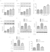
The expression levels of endothelial nitric oxide synthase (eNOS), vascular endothelial growth factor (VEGF) and hypoxia-inducible factor (HIF)-1α were increased by arnebin-1 in a phosphoinositide 3-kinase (PI3K)-dependent manner. (A–C) Human umbilical vein endothelial cells (HUVECs) were treated with arnebin-1 only at various concentrations (10−3, 10−2 and 10−1 µM) for 24 h. Cell lysates were subjected to western blot analysis. (A) Upper panel shows representative blots of the protein level of eNOS. Lower panel shows the quantification of the eNOS protein level. (B) Upper panel shows representative blots of the protein level of VEGF. Lower panel shows the quantification of the VEGF protein level. (C) The secretion level of VEGF in the culture supernatants was determined by ELISA. (D) Upper panel shows representative blots of the protein level of HIF-1α. Lower panel shows the quantification of the HIF-1α protein level. (E–H) HUVECs were treated with or without LY294002 for 1 h, and then stimulated with arnebin-1 in the presence or absence of VEGF for 24 h. (E) Upper panel shows representative blots of the protein level of HIF-1α. Lower panel shows the quantification of the HIF-1α protein level. (F) Upper panel shows representative blots of the protein level of eNOS. Lower panel shows the quantification of the eNOS protein level. (G) Upper panel shows representative blots of the protein level of VEGF. Lower panel shows the quantification of the VEGF protein level. (H) The secretion level of VEGF in the culture supernatants was determined by ELISA. Bars represent the means ± SEM. *P<0.05, **P<0.01 vs. control; #P<0.05, ##P<0.01 vs. Arnebin-1-treated groups. Control, vehicle-treated group.
Figure 4
Figure 4
The effect of arnebin-1 on the phosphoinositide 3-kinase (PI3K) pathway. (A–C) Human umbilical vein endothelial cells (HUVECs) were treated with arnebin-1 only, at various concentrations (10−3, 10−2 and 10−1 µM) for 24 h. Cell lysates were subjected to western blot analysis. (A) Upper panel shows representative blots of the protein level of total (t-)PI3K. Lower panel shows the quantification of the t-PI3K protein level. (B) Upper panel shows representative blots of the protein level of t-Akt. Lower panel shows the quantification of the t-Akt protein level. (C) Upper panel shows representative blots of the protein level of t-mTOR. Lower panel shows the quantification of the t-mTOR protein level. (D–F) HUVECs were treated with arnebin-1 only at various concentrations (10−3, 10−2 and 10−1 µM) for 2 h. Cell lysates were subjected to western blot analysis. (D) Upper panel shows representative blots of the protein level of phosphorylated (p-)PI3K and t-PI3K. Lower panel shows the quantification of the p-PI3K/t-PI3K protein level. (E) Upper panel shows representative blots of the protein level of p-Akt and t-Akt. Lower panel shows the quantification of the p-Akt/t-Akt protein level. (F) Upper panel shows representative blots of the protein level of p-mTOR and t-mTOR. Lower panel shows the quantification of the p-mTOR/t-mTOR protein level. Bars represent the means ± SEM. *P<0.05, **P<0.01 vs. control. Control, vehicle-treated group.
Figure 5
Figure 5
Hypoxia-inducible factor (HIF)-1α is essential for arnebin-1-induced (A) cell proliferation, (B and C) cell migration and (D–E) tube formation of human umbilical vein endothelial cells (HUVECs) in the presence of vascular endothelial growth factor (VEGF). HUVECs were treated with or without LY294002 (2 µM) for 1 h, and then stimulated with arnebin-1 (10−1 µM) in the presence or absence of VEGF (1 ng/ml) for 24 h. (A) Cell proliferation was assessed by MTT assay. HUVECs were treated with or without LY294002 (2 µM) for 1 h, and then stimulated with arnebin-1 (10−1 µM) in the presence or absence of VEGF (1 ng/ml) for 8 h. (B and C) Cell migration was assessed by Transwell assay. HUVECs were treated with or without LY294002 (2 µM) for 1 h, and then stimulated with arnebin-1 (10−1 µM) in the presence or absence of VEGF (1 ng/ml) for 12 h. (D–E) HUVECs were plated on Matrigel to form tubular structures. Bars represent the means ± SEM. *P<0.05, **P<0.01 vs. control; #P<0.05, ##P<0.01 vs. VEGF-treated group; and P<0.05 vs. VEGF + Arnebin-1-treated group. Control, vehicle-treated group.
Figure 6
Figure 6
Effects of arnebin-1 on the hypoxia-inducible factor (HIF)-1α, vascular endothelial growth factor (VEGF) and endothelial nitric oxide synthase (eNOS) expression levels in diabetic rats. (A–C) Effects of arnebin-1 on the protein expression levels of HIF-1α, VEGF and eNOS. (A) Upper panel shows representative blots of the protein level of HIF-1α. Lower panel shows the quantification of the HIF-1α protein level. (B) Upper panel shows representative blots of the protein level of eNOS. Lower panel shows the quantification of the eNOS protein level. (C) Upper panel shows representative blots of the protein level of VEGF. Lower panel shows the quantification of the VEGF protein level. Bars represent the means ± SEM. *P<0.05, **P<0.01 vs. non-diabetic rats; #P<0.05, ##P<0.01 vs. diabetic rats. n=6 for each group. D+V, diabetic rats treated with the vehicle; D+A, diabetic rats treated with arnebin-1.
Figure 7
Figure 7
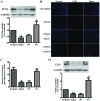
Effects of arnebin-1 on proliferation and neovascularization in diabetic rats. (A) Effects of arnebin-1 on the expression levels of proliferating cell nuclear antigen (PCNA), a nuclear cell proliferation marker. Upper panel shows representative blots of the protein level of PCNA. Lower panel shows the quantification of the PCNA protein level. (B) Effects of arnebin-1 on wound vascularity. Wound sections were stained with an anti-CD31 antibody and detected with Cy3 (red). Representative immunofluorescence images of wound samples on day 7 after treatment. Immunostaining for CD31-positive blood vessels (red) was performed to show vasculature in wounds, and nuclei (blue) were counterstained with Hoechst 33342. (C) Quantitative analysis of CD31-positive blood vessels in each section. Results are expressed as the number of vessels per high-power field. (D) Effects of arnebin-1 on the expression levels of CD31. Upper panel shows representative blots of the protein level of CD31. Lower panel shows the quantification of the CD31 protein level. Bars represent the means ± SEM. *P<0.05, **P<0.01 vs. non-diabetic rats; ##P<0.01 vs. diabetic rats. n=6 for each group. D+V, diabetic rats treated with the vehicle; D+A, diabetic rats treated with arnebin-1.
Figure 8
Figure 8
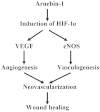
Schematic diagram of the mechanisms through which arnebin-1 promotes vascularization and wound healing. Arnebin-1 treatment leads to the accumulation of hypoxia-inducible factor (HIF)-1α, and the consequent upregulation of VEGF and endothelial nitric oxide synthase (eNOS). The expression of HIF-1α target genes in turn promotes neovascularization in diabetic wounds through angiogenesis and vasculogenesis.

Similar articles

Cited by

References

    1. Boulton AJ. The diabetic foot: Grand overview, epidemiology and pathogenesis. Diabetes Metab Res Rev. 2008;24(Suppl 1):S3–S6. doi: 10.1002/dmrr.833. - DOI - PubMed
    1. Boulton AJ, Vileikyte L, Ragnarson-Tennvall G, Apelqvist J. The global burden of diabetic foot disease. Lancet. 2005;366:1719–1724. doi: 10.1016/S0140-6736(05)67698-2. - DOI - PubMed
    1. Bartus CL, Margolis DJ. Reducing the incidence of foot ulceration and amputation in diabetes. Curr Diab Rep. 2004;4:413–418. doi: 10.1007/s11892-004-0049-x. - DOI - PubMed
    1. Macfarlane RM, Jeffcoate WJ. Factors contributing to the presentation of diabetic foot ulcers. Diabet Med. 1997;14:867–870. doi: 10.1002/(SICI)1096-9136(199710)14:10<867::AID-DIA475>3.0.CO;2-L. - DOI - PubMed
    1. Ruffieux P, Hommel L, Saurat JH. Long-term assessment of chronic leg ulcer treatment by autologous skin grafts. Dermatology. 1997;195:77–80. doi: 10.1159/000245698. - DOI - PubMed

Publication types

MeSH terms

Substances