. 2015 Sep 24;126(13):1585-94.
doi: 10.1182/blood-2015-03-635227. Epub 2015 Jul 22.

PKR inhibits the DNA damage response, and is associated with poor survival in AML and accelerated leukemia in NHD13 mice

Affiliations

PKR inhibits the DNA damage response, and is associated with poor survival in AML and accelerated leukemia in NHD13 mice

Xiaodong Cheng et al. Blood. .

Abstract

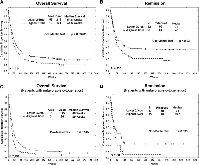
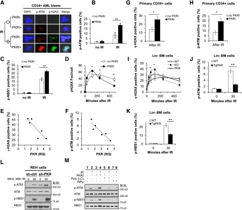
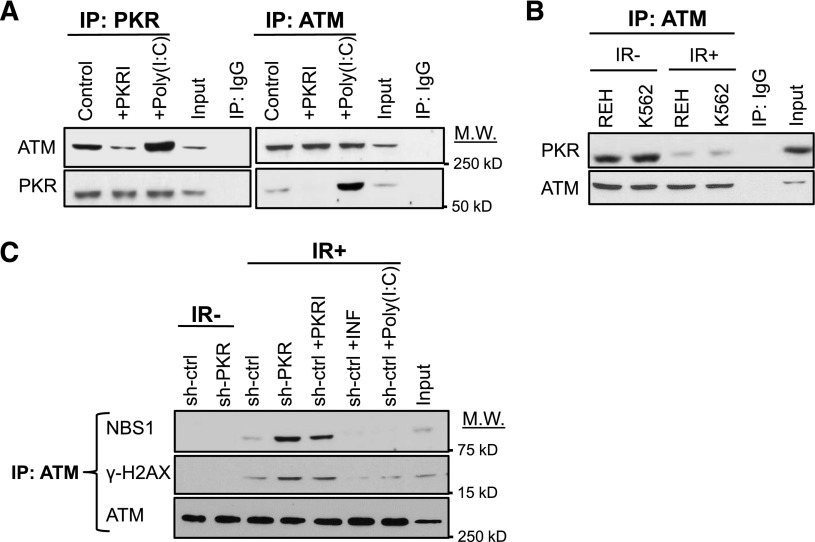
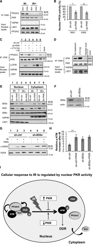
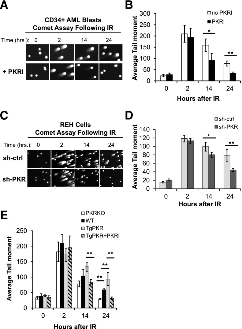
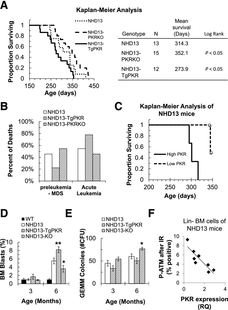
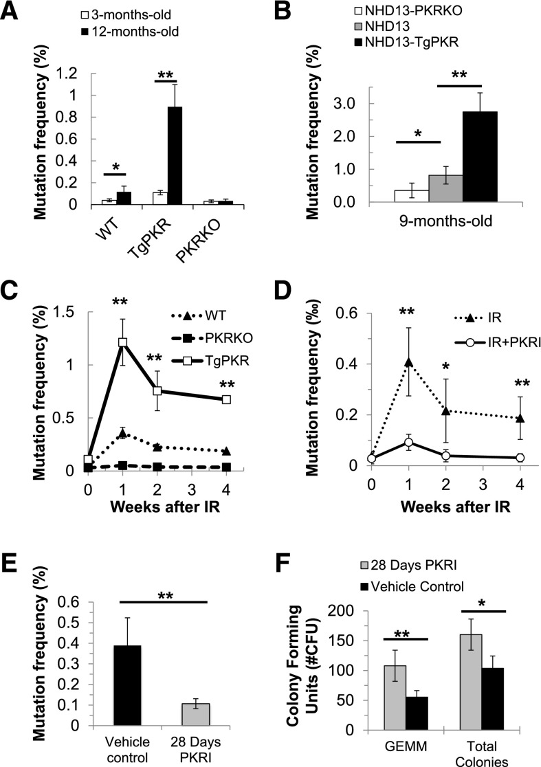
Increased expression of the interferon-inducible double-stranded RNA-activated protein kinase (PKR) has been reported in acute leukemia and solid tumors, but the role of PKR has been unclear. Now, our results indicate that high PKR expression in CD34(+) cells of acute myeloid leukemia (AML) patients correlates with worse survival and shortened remission duration. Significantly, we find that PKR has a novel and previously unrecognized nuclear function to inhibit DNA damage response signaling and double-strand break repair. Nuclear PKR antagonizes ataxia-telangiectasia mutated (ATM) activation by a mechanism dependent on protein phosphatase 2A activity. Thus, inhibition of PKR expression or activity promotes ATM activation, γ-H2AX formation, and phosphorylation of NBS1 following ionizing irradiation. PKR transgenic but not PKR null mice demonstrate a mutator phenotype characterized by radiation-induced and age-associated genomic instability that was partially reversed by short-term pharmacologic PKR inhibition. Furthermore, the age-associated accumulation of somatic mutations that occurs in the Nup98-HOXD13 (NHD13) mouse model of leukemia progression was significantly elevated by co-expression of a PKR transgene, whereas knockout of PKR expression or pharmacologic inhibition of PKR activity reduced the frequency of spontaneous mutations in vivo. Thus, PKR cooperated with the NHD13 transgene to accelerate leukemia progression and shorten survival. Taken together, these results indicate that increased nuclear PKR has an oncogenic function that promotes the accumulation of potentially deleterious mutations. Thus, PKR inhibition may be a therapeutically useful strategy to prevent leukemia progression or relapse, and improve clinical outcomes.


Figures

Figure 1
Figure 1
Increased PKR expression is associated with poor survival and shortened remission for acute leukemia patients. RPPA of CD34+ cells collected from PB and BM of 414 newly diagnosed AML patients was used to classify patients into 2 groups based on PKR expression level (highest one-third vs lowest two-third expression). Kaplan-Meier analysis was used to measure: (A) OS probability of all patients (n = 414), (B) remission duration for those patients who achieved complete remission (n = 230), (C) survival probability for those patients with unfavorable cytogenetics (n = 195), and (D) remission duration of patients with unfavorable cytogenetics (n = 80). Median (weeks) and P value for each group were calculated by Cox proportional hazard modeling (ie, Cox-Mantel test).
Figure 2
Figure 2
PKR inhibits DDR signaling in primary CD34+ AML cells, primary murine Lin BM cells, and human leukemic cell lines. (A) p(Ser1981)-ATM (p-ATM) and γ-H2AX were evaluated in CD34+ AML blast cells isolated from the PB and BM of 6 AML patients by IF microscopy (×63). Treatment with 0.5 μM PKRI for 8 hours increased p-ATM and γ-H2AX staining, both 30 minutes after 5 Gy IR. Images are of CD34+ cells from a single AML patient sample that are representative of results from 6 AML patients. (B-C) The percent of primary CD34+ AML blasts cells positive for p-ATM (B) and p(Ser343)-NBS1 (p-NBS1) (C) after 5 Gy IR was increased by 8 hours of treatment with 0.5 μM PKRI as measured by flow cytometry. Results are an average of CD34+ AML cells from 6 patients. (D) At the indicated times following 5 Gy IR treatment, primary CD34+ AML cells treated with PKRI displayed an increased rate and extent of γ-H2AX foci formation following IR. The number of γ-H2AX foci per cell was counted by IF microscopy of 30 randomly selected cells for each AML patient sample and the averages graphed. (E-F) Quantitative real-time polymerase chain reaction was used to measure PKR expression in CD34+ AML blasts from 6 patients relative to healthy donor CD34+ BM cells. The percent of γ-H2AX (E) or p-ATM positive (F) cells 30 minutes after 5 Gy IR was measured by flow cytometry and plotted against the relative quantity (RQ) of PKR expression for each of the 6 AML patient samples. (G-H) Treatment of primary human CD34+ cells isolated from BM of healthy donors with 0.5 μM PKRI for 8 hours increased the percentage of cells positive for γ-H2AX (G) and p-ATM (H) after 5 Gy IR as measured using flow cytometry. Results are an average of 3 independent experiments. (I) Lin BM cells were isolated from WT, PKRKO, or TgPKR mice and irradiated with 5 Gy. At the indicated times after IR, the percent of cells positive for γ-H2AX was measured by flow cytometry. (J) p-ATM and (K) p-NBS1 were decreased in Lin cells from BM of TgPKR mice compared with cells from WT mice. (L) Western blotting of REH cells demonstrates that p-ATM and p-NBS1 are increased in cells with reduced PKR expression by shRNA knockdown (sh-PKR) compared with cells expressing a control shRNA (sh-ctrl), both under normal growth conditions and 30 minutes after treatment with 5 Gy IR. (M) REH cells pretreated with PKRI (0.5 μM for 8 hours), IFN-γ (10 ng/mL for 24 hours), or poly(I:C) (5 μg/mL for 6 hours) demonstrate that increased PKR expression and activity decreased p-ATM and p-NBS1 30 minutes after IR. *P < .05; **P < .01. DAPI, 4,6 diamidino-2-phenylindole; M.W., molecular weight.
Figure 3
Figure 3
Activated PKR associates with ATM and inhibits ATM activation. (A) Western blotting after reciprocal co-IP of ATM and PKR from lysates of REH cells treated with either 0.5 μM PKRI for 8 hours or 5 μg/mL poly (I:C) for 6 hours demonstrates that PKR activity enhances PKR-ATM association. Input is 10% of total lysate used in the co-IP. Nonspecific IgG antibody was used as a negative co-IP control. (B) Western blotting indicates that co-IP of ATM and PKR is decreased at 30 minutes after 5 Gy IR. (C) co-IP of ATM with NBS1 or γ-H2AX is increased in REH cells by inhibition of PKR expression or activity, whereas cells treated with 10 ng/mL IFN-γ or 5 μg/mL poly(I:C) to increase PKR expression/activity have a decreased co-IP of ATM with either NBS1 or γ-H2AX.
Figure 4
Figure 4
Nuclear PKR activates PP2A to inhibit ATM phosphorylation. (A) PKR co-IPs with PP2A from the nuclear fraction of REH and K562 cell lysates but this association is significantly reduced 30 minutes after exposure to 5 Gy IR (IR+). Western blotting for HSP90 (cytoplasm) and LSD1 (nucleus) was performed as a subcellular fractionation control. Input is 10% of total REH nuclear lysate used in the co-IP. (B) PP2A activity in the nuclear fraction of leukemia cell lines is reduced in cells with decreased PKR expression (sh-PKR) compared with cells transfected with a control shRNA (sh-ctrl). (C) co-IP of PP2A and ATM from the nuclear lysate of REH sh-ctrl and REH sh-PKR cells 30 minutes after treatment with 5 Gy IR and/or pretreatment with 2.5 μM of the PP2A activator FTY720 for 8 hours revealed that PP2A and ATM association is decreased after irradiation, decreased by reduced PKR expression, but increased by FTY720 treatment. In addition, p-ATM is correspondingly increased when ATM-PP2A nuclear association is decreased. Input is 10% of total REH sh-ctrl nuclear lysate used in the co-IP. (D) co-IP of PKR with ATM from the nuclear fraction of REH cells is increased by treatment with 2.5 μM of the PP2A activator FTY720 for 8 hours, whereas PKR-ATM association is decreased by treatment with 1 μM of the PP2A inhibitor OA for 8 hours. Vertical dashed line indicates a repositioned gel lane. (E) Expression of PP2A-B55α and PP2A-B56γ subunits in the cytoplasm and nucleus of REH cells were detected by western blotting. Knockdown of PKR decreased nuclear B55α and increased cytoplasmic B55α both before and after 5 Gy IR. (F) Western blotting demonstrates that B55α expression in REH cells was decreased by B55α-specific shRNA (sh-B55α) compared with control shRNA (sh-ctrl) cells. (G) γ-H2AX formation 30 minutes after 5 Gy IR was measured by western blotting. Pretreatment of cells with 0.5 μM PKRI for 8 hours prior to IR promotes γ-H2AX formation in REH sh-ctrl cells but not REH sh-B55α cells with decreased B55α expression. (H) Flow cytometry after IR reveals that PKRI promotes increased p-ATM in control REH cells (sh-ctrl) but not B55α knockdown cells (sh-B55α). (I) Proposed model by which nuclear PKR mediates PP2A activity and DDR signaling following IR. In undamaged cells, nuclear PKR indirectly antagonizes ATM activation by promoting nuclear localization of the PP2A B55α regulatory subunit that increases nuclear PP2A phosphatase activity to inhibit ATM autophosphorylation. Following IR, PKR and PP2A no longer interact with the ATM complex, and the PP2A-B55α subunit is sequestered in the cytoplasm allowing ATM to be activated and initiate DDR signaling events. *P < .05; **P < .01.
Figure 5
Figure 5
Inhibition of PKR expression or activity promotes DNA DSB repair in hematopoietic cells. Neutral Comet assays were used to measure DNA DSB repair following 5 Gy IR. (A) Inhibition of PKR by treatment with 0.5 μM PKRI prior to and following IR promoted faster kinetics of DNA DSB repair in primary CD34+ AML cells. SYBR gold-stained Comets were visualized by microscopy (×40). A representative Comet assay of 1 patient sample is shown. (B) The average Comet Olive Tail Moment from 50 randomly chosen CD34+ AML cells in each of 6 patient samples was determined. (C) Representative Comet assay, and (D) calculation of the average Comet Tail Moment of 50 randomly chosen REH cells demonstrates reduced PKR expression (sh-PKR) increases the rate of DNA DSB repair compared with control (sh-ctrl) cells. (E) Lin BM cells were collected from WT, TgPKR, and PKRKO mice, and treated with 5 Gy IR. Comet Tail Moments were calculated from 50 randomly chosen cells for each genotype. Lin BM cells from PKRKO mice displayed more rapid kinetics of DNA DSB repair than WT cells. Compared with WT, TgPKR cells exhibit delayed DNA DSB repair that can be restored by treatment with 0.5 μM PKRI. *P < .05; **P < .01.
Figure 6
Figure 6
PKR expression cooperates with the NHD13 transgene to shorten survival and promote MDS evolution to acute leukemia in the NHD13 mouse. (A) NHD13 mice were crossed with mice expressing a PKR transgene specifically in hematopoietic cells (TgPKR) or PKRKO mice, to produce NHD13-TgPKR and NHD13-PKRKO mice. NHD13 (n = 12), NHD13-TgPKR (n = 12), and NHD13-PKRKO (n = 12) mice were aged until physical deterioration led to death or required euthanasia (defined as body condition score ≤2). (A) Kaplan–Meier analysis demonstrates that PKR expression cooperates with NHD13 to significantly shorten the survival of mice. (B) NHD13-TgPKR mice more frequently die of acute leukemia than NHD13 mice. Acute leukemia or MDS was determined by hematoxylin and eosin staining and flow cytometry of BM cells collected at time of death. (C) Kaplan–Meier analysis demonstrates superior survival in NHD13 mice with low-level PKR expression vs NHD13 mice with high-level PKR expression (RQ >2) relative to age-matched WT controls. PKR expression in PB mononuclear cells collected as NHD13 mice aged was measured by quantitative real-time polymerase chain reaction. Circles represent mice that were censored at 350 days. (D-E) At 3 and 6 months of age, BM was collected from WT, NHD13, NHD13-TgPKR, and NHD13-PKRKO mice for comparison by flow cytometry analysis. (D) BM blasts were measured using CD45+ expression and side scatter. NHD13-TgPKR mice had significantly increased BM blasts compared with NHD13 mice at 6 months, whereas in PKRKO mice, this was significantly reduced. (E) BM from NHD13-PKRKO mice had significantly increased CFU-GEMM activity compared with NHD13 or NHD13-TgPKR. (F) Lin BM cells were isolated from 8 NHD13 mice and PKR level determined by flow cytometry. Some 30 minutes after IR, p-ATM was measured by flow cytometry and plotted vs PKR expression to reveal that relative PKR expression was inversely proportional to p-ATM. Each point represents an individual mouse. *P < .05; **P < .01.
Figure 7
Figure 7
Inhibition of PKR protects against mutations induced by age, IR, or a potent oncogene. The mutation frequency was measured serially in PB reticulocytes using the in vivo PIG-A mutation assay that detects the loss of the glycosylphosphatidylinositol-linked proteins like CD24 on the surface of reticulocytes by flow cytometry. The PIG-A mutation frequency was calculated as the percentage of CD24 reticulocytes/total reticulocytes analyzed. (A) PB from unperturbed young (3-month-old) vs old (12-month-old) WT, PKRKO, and TgPKR mice was obtained, and reticulocytes isolated and analyzed by flow cytometry for the presence or absence of CD24. Increased PKR expression in TgPKR hematopoietic cells promotes an age-associated increase in the frequency of PIG-A mutation compared with WT or PKRKO. (B) NHD13-TgPKR mice, aged 9 months, have a significantly increased frequency of PIG-A mutation compared with NHD13 or NHD13-PKRKO mice. (C) Increased PKR expression promotes a significant increase in the frequency of PIG-A mutations following 5 Gy IR exposure. (D) PKRI treatment reduces the frequency of PIG-A somatic mutation in PB cells of mice following IR. WT mice were injected intraperitoneally with either 200 μg/kg PKRI or PBS (5 mice in each treatment group) and administered a single sublethal dose of IR (5 Gy). After IR, mice received either 200 μg/kg PKRI or PBS every 12 hours for 4 weeks. (E-F) NHD13 mice that were 4 months old were implanted with Alzet osmotic pumps (#1004) filled with either PKRI (1.5 mg/mL in PBS:dimethylsulfoxide) or vehicle control (PBS:dimethylsulfoxide). After 28 days, mice were euthanized to collect blood and BM. (E) The PIG-A mutation frequency in PB of NHD13 mice was significantly reduced by 28-day continuous PKRI treatment. (F) BM of NHD13 mice that received continuous PKRI treatment had significantly greater CFU-GEMM and total CFU activity. *P < .05; **P < .01.

Comment in

Similar articles

Cited by

References

    1. Bennett RL, Blalock WL, Abtahi DM, Pan Y, Moyer SA, May WS. RAX, the PKR activator, sensitizes cells to inflammatory cytokines, serum withdrawal, chemotherapy, and viral infection. Blood. 2006;108(3):821–829. - PMC - PubMed
    1. Ito T, Jagus R, May WS. Interleukin 3 stimulates protein synthesis by regulating double-stranded RNA-dependent protein kinase. Proc Natl Acad Sci USA. 1994;91(16):7455–7459. - PMC - PubMed
    1. Liu X, Bennett RL, Cheng X, Byrne M, Reinhard MK, May WS., Jr PKR regulates proliferation, differentiation, and survival of murine hematopoietic stem/progenitor cells. Blood. 2013;121(17):3364–3374. - PMC - PubMed
    1. Bennett RL, Pan Y, Christian J, Hui T, May WS., Jr The RAX/PACT-PKR stress response pathway promotes p53 sumoylation and activation, leading to G₁ arrest. Cell Cycle. 2012;11(2):407–417. - PMC - PubMed
    1. Cuddihy AR, Li S, Tam NW, et al. Double-stranded-RNA-activated protein kinase PKR enhances transcriptional activation by tumor suppressor p53. Mol Cell Biol. 1999;19(4):2475–2484. - PMC - PubMed

Publication types

MeSH terms