. 2015 Sep 15;26(18):3215-28.
doi: 10.1091/mbc.E14-07-1246. Epub 2015 Jul 22.

Positive and negative regulation by SLP-76/ADAP and Pyk2 of chemokine-stimulated T-lymphocyte adhesion mediated by integrin α4β1

Affiliations

Positive and negative regulation by SLP-76/ADAP and Pyk2 of chemokine-stimulated T-lymphocyte adhesion mediated by integrin α4β1

Ana Dios-Esponera et al. Mol Biol Cell. .

Abstract

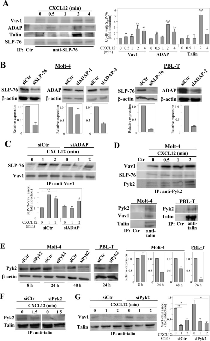
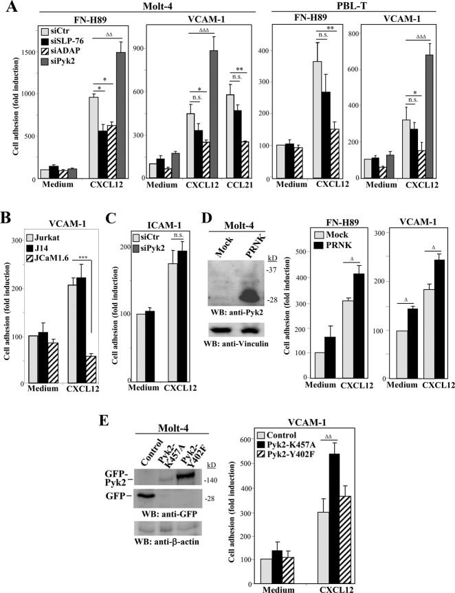
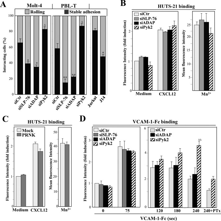
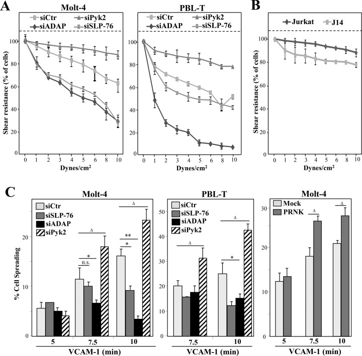
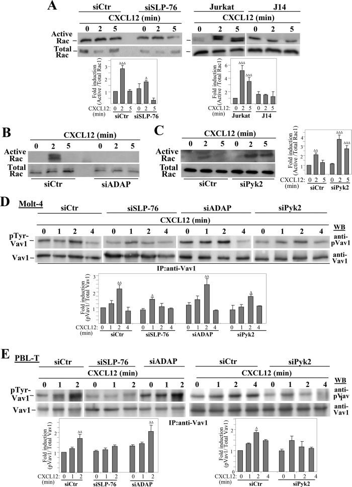
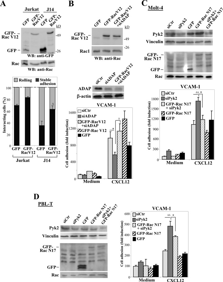
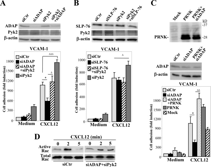
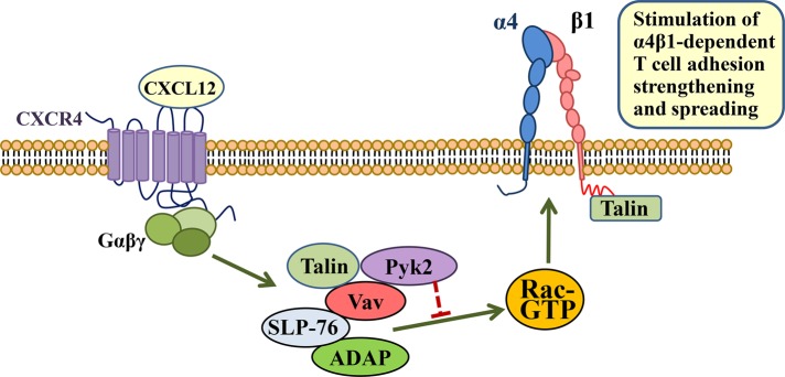
Stimulation by chemokines of integrin α4β1-dependent T-lymphocyte adhesion is a crucial step for lymphocyte trafficking. The adaptor Vav1 is required for chemokine-activated T-cell adhesion mediated by α4β1. Conceivably, proteins associating with Vav1 could potentially modulate this adhesion. Correlating with activation by the chemokine CXCL12 of T-lymphocyte attachment to α4β1 ligands, a transient stimulation in the association of Vav1 with SLP-76, Pyk2, and ADAP was observed. Using T-cells depleted for SLP-76, ADAP, or Pyk2, or expressing Pyk2 kinase-inactive forms, we show that SLP-76 and ADAP stimulate chemokine-activated, α4β1-mediated adhesion, whereas Pyk2 opposes T-cell attachment. While CXCL12-promoted generation of high-affinity α4β1 is independent of SLP-76, ADAP, and Pyk2, the strength of α4β1-VCAM-1 interaction and cell spreading on VCAM-1 are targets of regulation by these three proteins. GTPase assays, expression of activated or dominant-negative Rac1, or combined ADAP and Pyk2 silencing indicated that Rac1 activation by CXCL12 is a common mediator response in SLP-76-, ADAP-, and Pyk2-regulated cell adhesion involving α4β1. Our data strongly suggest that chemokine-stimulated associations between Vav1, SLP-76, and ADAP facilitate Rac1 activation and α4β1-mediated adhesion, whereas Pyk2 opposes this adhesion by limiting Rac1 activation.


Figures

FIGURE 1:
FIGURE 1:
Chemokine-stimulated associations between Vav1, SLP-76, ADAP, and Pyk2. (A) Left, Molt-4 T-cells incubated for the indicated times with CXCL12 were subjected to immunoprecipitation with anti–SLP-76 followed by immunoblotting with antibodies to the indicated proteins. Right, bars represent densitometric quantification of gel bands showing the mean ± SD of six independent experiments (ΔΔΔ, p < 0.001; ΔΔ, p < 0.01; Δ, p < 0.05). (B) Top, cells were transfected with SLP-76, ADAP, or control siRNA, and expression of SLP-76 and ADAP was analyzed by immunoblotting. Control loading is shown by blotting with anti–β-actin antibodies. Bottom, densitometric quantification of gel bands showing the mean ± SD of four (Molt-4) or three (PBL-T) independent experiments. (C) CXCL12-incubated control or ADAP siRNA Molt-4 transfectants were assayed by immunoprecipitation with anti-Vav1 antibodies, followed by immunoblotting with antibodies to the proteins shown (ΔΔ, p < 0.01; Δ, p <0.05). (D) Cells were incubated in the absence or presence of CXCL12, and subsequently subjected to immunoprecipitation and Western blotting. (E) Left, Cells were transfected with Pyk2 or control siRNA, and transfectants were assayed by Western blotting at the indicated times. Right, densitometric analyses of gel bands showing the mean ± SD of three independent experiments. (F and G) Control or Pyk2 siRNA transfectants were subjected to immunoprecipitation with anti-talin antibodies, followed by immunoblotting with antibodies to the shown proteins. Talin-Vav1 coprecipitation was significantly diminished (**, p < 0.001; *, p < 0.05; n = 4).
FIGURE 2:
FIGURE 2:
Chemokine-stimulated static adhesion of SLP-76, ADAP, or Pyk2 transfectants to α4β1 ligands. (A) Control, SLP-76–, ADAP-, or Pyk2-knockdown Molt-4 or PBL-T transfectants were subjected to adhesion assays to FN-H89 or VCAM-1 in the absence (Medium) or presence of coimmobilized CXCL12 or CCL21 (n = 3–5). (B) Parental Jurkat, J14, and JCaM1.6 cells were tested in adhesion assays to VCAM-1 as in A (n = 2). (C) Molt-4 cells were transfected with control or Pyk2 siRNA and transfectants tested in adhesion assays to ICAM-1 coimmobilized with or without CXCL12 (n = 3). (D) Cells were transfected with empty (Mock) or PRNK vectors, and transfectants were tested by Western blotting for PRNK expression (left) or in adhesion assays (middle and right) (n = 4). (E) Cells were transfected with control GFP vector or with the indicated GFP-fused Pyk2 mutants, and transfectants were subjected to immunoblotting or to adhesion assays (n = 4). Adhesions were significantly inhibited (***, p < 0.001; **, p < 0.01; *, p < 0.05) or significantly stimulated (ΔΔΔ, p < 0.001; ΔΔ, p < 0.01; Δ, p <0.05) (n.s., nonsignificant).
FIGURE 3:
FIGURE 3:
Flow-chamber adhesion assays and determination of high-affinity α4β1 expression in SLP-76–, ADAP-, or Pyk2-depleted T-cells. (A) Control, SLP-76, ADAP, or Pyk2 siRNA transfectants or Jurkat or J14 cells were perfused in flow chambers coated with VCAM-1 coimmobilized with CXCL12 and analyzed for rolling and stable cell arrest (n = 3–4). Data are presented as mean ± SD of cell percentages from the total cell population. Adhesions were significantly inhibited or stimulated in comparison with those of control siRNA transfectants or parental Jurkat cells, *p < 0.05 or Δp < 0.05, respectively. (B and C) The indicated siRNA Molt-4 transfectants or cells transfected with PRNK or empty vector were tested by flow cytometry for HUTS-21 mAb binding after stimulation with CXCL12 or Mn2+. (D) Following exposure to CXCL12 for 20 s, transfectants were analyzed by flow cytometry for VCAM-1-Fc binding after the indicated times. PTx denotes cells preincubated with pertussis toxin.
FIGURE 4:
FIGURE 4:
Analyses of T-cell adhesion strengthening and spreading on VCAM-1. The indicated siRNA transfectants (A) or Jurkat and J14 cells (B), preattached onto coimmobilized VCAM-1 and CXCL12 in flow chambers, were subjected to cell detachment after sequential increases of shear stress. Data show mean ± SD of cell percentages from the initial number of bound cells remaining attached at the indicated shear stresses (A, n = 4; B, n = 3). (C) Molt-4 and PBL-T were transfected with the indicated siRNA or vectors, and transfectants were allowed to attach to VCAM-1 immobilized with CXCL12. Spreading was evaluated from Nomarski images at the indicated times. Percentage of cell spreading was determined from cells (2500–3000) from different fields of view. Spreading was significantly inhibited (**, p < 0.01; *, p < 0.05) or stimulated (Δ, p < 0.05) compared with control siRNA or mock transfectants (n = 3). n.s., nonsignificant.
FIGURE 5:
FIGURE 5:
Analyses of Rac1 activation and Vav1 tyrosine phosphorylation in SLP-76–, ADAP-, or Pyk2-deficient T-cells. Control, SLP-76, ADAP, or Pyk2 siRNA transfectants in Molt-4 cells, or Jurkat and J14 cells (A–C) were stimulated with CXCL12 and subjected to GTPase assays to detect Rac1 activation. Activation was significantly increased compared with untreated cells (ΔΔΔ, p < 0.001; ΔΔ, p < 0.01; Δ, p < 0.05; A, n = 7; B and C, n = 3). The indicated Molt-4 (D) or PBL-T (E) siRNA transfectants incubated with or without CXCL12 for the indicated times were analyzed by immunoprecipitation and Western blotting. Vav1 tyrosine phosphorylation was significantly up-regulated compared with untreated cells (ΔΔ, p < 0.01; Δ, p < 0.05; D, n = 6; E, left, n = 2; E, right, n = 3).
FIGURE 6:
FIGURE 6:
Rac1 involvement in SLP-76, ADAP, and Pyk2 regulation of CXCL12-activated T-cell adhesion dependent on α4β1. (A) Cells were transfected with GFP or GFP-Rac V12 vectors and were analyzed by immunoblotting (top) or were subjected to flow-chamber adhesion assays to VCAM-1 coimmobilized with CXCL12 (bottom). Adhesion was significantly rescued in comparison with J14-GFP transfectants (Δ, p < 0.05; n = 3). (B) Molt-4 cells were transfected with the indicated siRNA and GFP vector combinations and were analyzed by Western blotting (top) or in static adhesion assays to VCAM-1 immobilized with or without CXCL12 (bottom). Adhesion was significantly inhibited (*, p < 0.05) or recovered (Δ, p < 0.05). (C and D) The indicated Molt-4 or PBL-T transfectants were tested by immunoblotting or were subjected to adhesion assays to VCAM-1 immobilized with or without CXCL12. Adhesion was significantly stimulated (ΔΔ, p < 0.01) or inhibited (*, p < 0.05) (B–D, n = 3).
FIGURE 7:
FIGURE 7:
Analysis of functional connections between ADAP and Pyk2 in CXCL12-stimulated adhesion mediated by α4β1. (A–C) Molt-4 cells were transfected with the indicated individual or combined siRNA and/or vectors, and transfectants were analyzed by immunoblotting or in adhesion assays to VCAM-1 immobilized with or without CXCL12. Adhesions were significantly inhibited (*p < 0.05) or significantly stimulated or rescued (ΔΔΔ, p < 0.001; ΔΔ, p < 0.01; or Δ, p < 0.05) (A, n = 4; B and C, n = 3). (D) The indicated transfectants were tested in GTPase assays to detect active Rac.
FIGURE 8:
FIGURE 8:
Model for regulation by Vav1-associated proteins of chemokine-stimulated, α4β1-dependent T-cell adhesion. Rac1 activation by CXCL12 is a well-known event for stimulation of the adhesion strength and spreading mediated by α4β1 in T-cells. A transient complex formed by Vav1, SLP-76, ADAP, and Pyk2 regulates this adhesion. While Vav1-SLP-76-ADAP stimulates chemokine-promoted Rac1 activation (green arrow), the presence of Pyk2 in the complex opposes this activation (red blocking symbol). The result of these opposing actions is a net Rac activation and up-regulation of T-cell adhesion strengthening and spreading dependent on α4β1. Talin can also be found in this complex, perhaps as an independent pool from β1-associated talin. Its role in the assembly and function of this complex has not been addressed in the present study.

Similar articles

Cited by

References

    1. Avraham H, Park SY, Schinkmann K, Avraham S. RAFTK/Pyk2-mediated cellular signalling. Cell Signal. 2000;12:123–133. - PubMed
    1. Avraham S, London R, Fu Y, Ota S, Hiregowdara D, Li J, Jiang S, Pasztor LM, White RA, Groopman JE, et al. Identification and characterization of a novel related adhesion focal tyrosine kinase (RAFTK) from megakaryocytes and brain. J Biol Chem. 1995;270:27742–27751. - PubMed
    1. Baker RG, Hsu CJ, Lee D, Jordan MS, Maltzman JS, Hammer DA, Baumgart T, Koretzky GA. The adapter protein SLP-76 mediates “outside-in” integrin signaling and function in T cells. Mol Cell Biol. 2009;29:5578–5589. - PMC - PubMed
    1. Barda-Saad M, Shirasu N, Pauker MH, Hassan N, Perl O, Balbo A, Yamaguchi H, Houtman JC, Appella E, Schuck P, Samelson LE. Cooperative interactions at the SLP-76 complex are critical for actin polymerization. EMBO J. 2010;29:2315–2328. - PMC - PubMed
    1. Beinke S, Phee H, Clingan JM, Schlessinger J, Matloubian M, Weiss A. Proline-rich tyrosine kinase-2 is critical for CD8 T-cell short-lived effector fate. Proc Natl Acad Sci USA. 2010;107:16234–16239. - PMC - PubMed

Publication types

MeSH terms