. 2015 Jul 23:10:31.
doi: 10.1186/s13024-015-0028-5.

Synaptotagmins interact with APP and promote Aβ generation

Affiliations

Synaptotagmins interact with APP and promote Aβ generation

Vivek Gautam et al. Mol Neurodegener. .

Abstract

Background: Accumulation of the β-amyloid peptide (Aβ) is a major pathological hallmark of Alzheimer's disease (AD). Recent studies have shown that synaptic Aβ toxicity may directly impair synaptic function. However, proteins regulating Aβ generation at the synapse have not been characterized. Here, we sought to identify synaptic proteins that interact with the extracellular domain of APP and regulate Aβ generation.

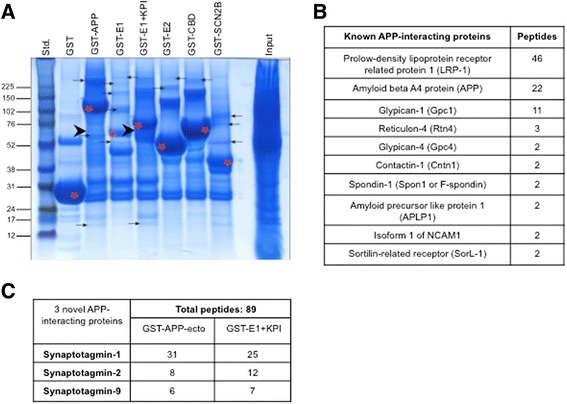
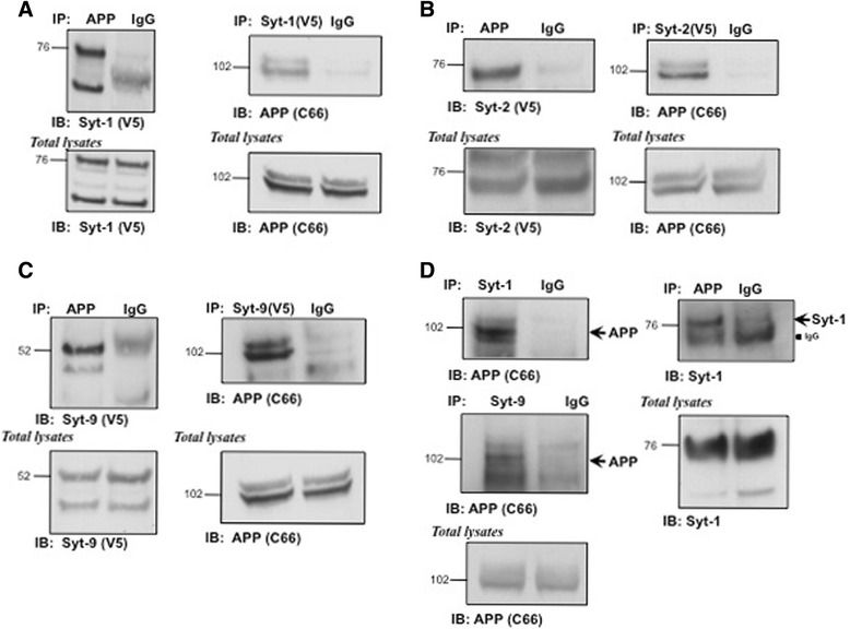
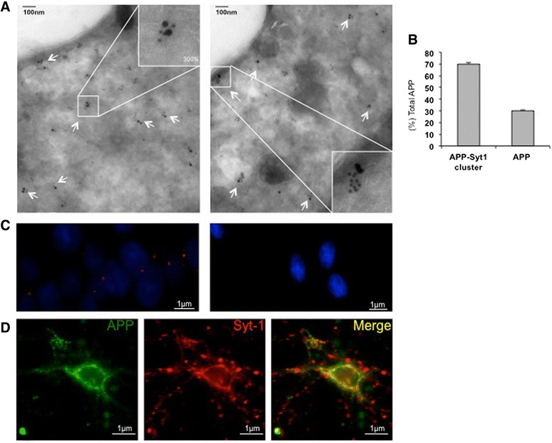
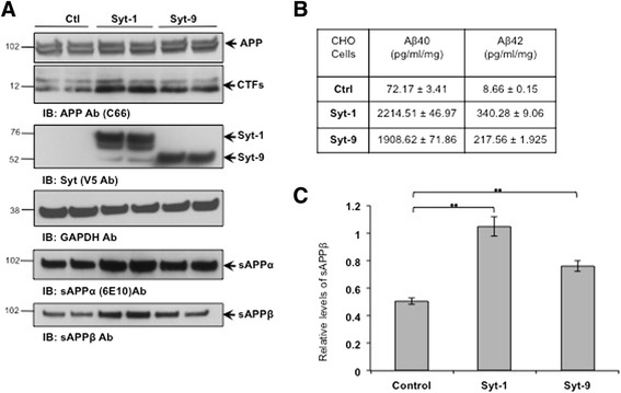
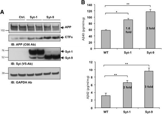
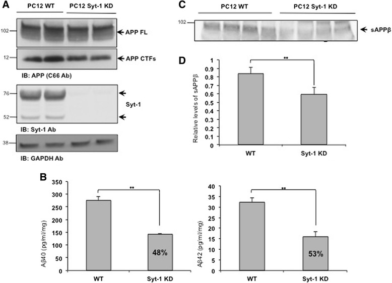
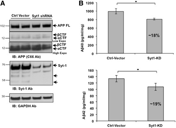
Results: Affinity purification-coupled mass spectrometry identified members of the Synaptotagmin (Syt) family as novel interacting proteins with the APP ectodomain in mouse brains. Syt-1, -2 and -9 interacted with APP in cells and in mouse brains in vivo. Using a GST pull-down approach, we have further demonstrated that the Syt interaction site lies in the 108 amino acids linker region between the E1 and KPI domains of APP. Stable overexpression of Syt-1 or Syt-9 with APP in CHO and rat pheochromocytoma cells (PC12) significantly increased APP-CTF and sAPP levels, with a 2 to 3 fold increase in secreted Aβ levels in PC12 cells. Moreover, using a stable knockdown approach to reduce the expression of endogenous Syt-1 in PC12 cells, we have observed a ~ 50% reduction in secreted Aβ generation. APP processing also decreased in these cells, shown by lower CTF levels. Lentiviral-mediated knock down of endogenous Syt-1 in mouse primary neurons also led to a significant reduction in both Aβ40 and Aβ42 generation. As secreted sAPPβ levels were significantly reduced in PC12 cells lacking Syt-1 expression, our results suggest that Syt-1 regulates Aβ generation by modulating BACE1-mediated cleavage of APP.

Conclusion: Altogether, our data identify the synaptic vesicle proteins Syt-1 and 9 as novel APP-interacting proteins that promote Aβ generation and thus may play an important role in the pathogenesis of AD.


Figures

Fig. 1
Fig. 1
Mass spectrometry-based identification of novel APP-interacting proteins. a Colloidal Blue-stained gel of APP-interacting proteins from mouse forebrain extracts. The proteins were pulled down with GST-tagged APP-ectodomain regions. Red asterisks denote purified GST-tagged APP-ectodomain fragments while arrows point to the bands that were excised and subjected to MS-based analysis for protein identification. Arrowheads denote bands containing Syt peptides. b MS-based confirmation of previously identified APP ectodomain-interacting proteins. c MS-based identification of Syt-1, Syt-2 and Syt-9 peptides. Peptides from all three Syts were pulled down by GST-APP and GST-E1 + KPI. Other GST-tagged APP fragments did not pull down Syt peptides (not shown)
Fig. 2
Fig. 2
APP interacts with Syt-1, −2 and −9 in vitro and in vivo. a Western blot analysis of APP and Syt-1 co-immunoprecipitates. CHO cells stably coexpressing APP and V5-tagged Syt-1 were subjected to immunoprecipitation using specific APP (C66) or V5-tag antibodies. Syt-1 or APP specific bands were identified in the immunoprecipitates but were not observed in the control IgG pull-down (n = 3 for each condition). b Co-immunoprecipitation of APP and Syt-2. APP or Syt-2 were immunoprecipitated from CHO cells expressing Syt-2 (V5-tag) and probed with anti-V5 or APP (C66) antibodies. APP specific bands were identified in the fraction immunoprecipitated with anti V5 antibody (Syt-2) while Syt-2 specific bands were identified in the APP pull-down (n = 3 for each condition). c Western blot analysis showing co-immunoprecipitation of APP with Syt-9. CHO cells stably co-expressing APP and V5-tagged Syt-9 were subjected to immunoprecipitation using specific APP (C66) or V5-tag antibodies. Syt-9 or APP specific bands were identified in the immunoprecipitates but were not observed in the control IgG pull-down (n = 3 for each condition). d Western blot analysis of APP and Syt-1 or Syt-9 co-immunoprecipitates from adult mouse forebrain. Specific antibodies against endogenous Syt-1 or Syt-9 were used to immunoprecipitate Syt-1 or Syt-9 and probed with an anti-APP antibody. Western blot shows APP-specific staining in both Syt-1 and Syt-9 immunoprecipitates as compared to the IgG controls. In a reverse co-immunoprecipitation assay, the APP-specific antibody co-immunoprecipiated Syt-1 (n = 2 for each condition)
Fig. 3
Fig. 3
Syt-1 is a physiological interactor of APP. a Electron microscopic (EM) images of ultra-thin cryosections of naïve PC12 cells show double immunogold labeling of endogenous APP (15 nm) with Syt-1 (10 nm). White arrows depict co-localization of APP with Syt-1 while the inserts show 300 % magnification of APP in close association with Syt-1. Analysis was based on three independent sets of experiments. b Quantitative analysis of immunogold labeling of APP in cluster with Syt-1 compared to APP alone, as revealed by a. c In situ PLA shows interaction between endogenous APP and Syt-1 in naïve PC12 cells. Fluorescence red dots represent close association between APP and Syt-1 (left panel) as compared to the negative control (right panel). Images were taken at identical settings. Nuclear staining with DAPI is shown in blue. Analysis was based on three independent sets of experiments. d Immunofluorescence microscopy images of mouse primary neuronal cultures stained with APP and Syt-1 specific antibodies. Co-localization between APP (green) and Syt-1 (red) was observed in the cell body and in neurites. Images were obtained at identical settings. Analysis was based on three independent sets of experiments
Fig. 4
Fig. 4
APP interacts with Syt-1 and Syt-9 via its linker region between the E1 and KPI domain. a In vitro GST pull-down assay of Syt-1, −2 and −9. GST-tagged APP-ectodomain fragments were used to pull down V5-tagged Syt-1, −2 and −9 from stably expressing CHO cells. Western blot analysis with anti-V5 antibodies revealed specific interaction of Syt-1, −2 and −9 with both GST-APP-ectodomain and GST-E1 + KPI fragments. Analysis was based on 4 different sets of experiments. b In vitro GST pull-down assay showing interaction of Syt-1 and Syt-9 with the linker region of APP between the E1 and KPI domain. The GST-tagged APP-289 fragment (containing the linker region but not the KPI domain) pulled down both Syt-1 and Syt-9 while the GST-E1 domain did not. Analysis was based on three different sets of experiments. c Co-immunoprecipitation of the APP695 isoform with Syt-1 and Syt-9. CHO cells stably expressing APP695 were transfected with V5-tagged Syt-1 or Syt-9. Western blot analysis shows specific co-immunoprecipitation of APP695 with V5-tagged Syt-1 and Syt-9 (n = 3 for each condition). d In vitro GST pull-down assay showing that the Syt-1 N-terminal region interacts with APP. Purified GST-Syt1 N-terminal region pulled down full-length APP while no interaction was observed with control GST. Bottom panel shows separate colloidal blue stained gel of purified GST and GST-Syt1 N-terminal purified proteins used for the APP pull-down assay. Analysis was based on four different sets of experiments
Fig. 5
Fig. 5
Syt-1 and Syt-9 modulate APP processing and Aβ levels in CHO cells. a Western blot analysis of CHO cells stably co-expressing Syt-1 or Syt-9 with APP shows a significant increase in APP-CTFs, sAPPα and sAPPβ levels as compared to the control CHO-APP cells. Syt-1 and Syt-9 expression was confirmed using anti-V5 tag antibodies and a GAPDH antibody was used for equal protein loading. b Sandwich ELISA from the conditioned media indicates a strong increase in Aβ40 and Aβ42 release from cells expressing Syt-1 or Syt-9 as compared to the control CHO-APP cells. c Quantitative analysis of total sAPPβ levels in the conditioned media of CHO cells co-expressing Syt-1 or Syt-9 with APP as compared to only APP expression (student t test; **, p < 0.01; n = 4 for each condition)
Fig. 6
Fig. 6
Expression of Syt-1 and Syt-9 in PC12 cells increases endogenous APP-CTF and Aβ levels. a Western blot analysis shows that stable expression of Syt-1 or Syt-9 in PC12 cells increases endogenous APP-CTF levels. Bottom panel shows expression of Syt-1 and Syt-9 and a GAPDH antibody was used for equal protein loading. b Quantitative analysis of sandwich ELISA from the conditioned media of PC12 cells co-expressing Syt-1 or Syt-9 with APP. Expression of Syt-1 or Syt-9 in PC12 cells causes a robust increase in secreted Aβ40 and Aβ42 levels as compared to the control APP-expressing cells (student t test; *, p < 0.05; **, p < 0.01; n = 4 for each condition)
Fig. 7
Fig. 7
Stable knockdown of Syt-1 reduces endogenous APP-CTF, Aβ and sAPPβ levels in PC12 cells. a Stable knockdown of Syt-1 in PC12 cells reduces endogenous APP-CTF levels as revealed by Western blot analysis. Knock down of Syt-1 was confirmed with a Syt-1 specific antibody (Synaptic Systems) and a GAPDH antibody was used for equal protein loading. b Quantitative analysis of sandwich ELISA shows lower Aβ40 and Aβ42 levels in the conditioned media of the PC12 cells with stable Syt-1 KD as compared to the WT cells (student t test; **, p < 0.01; n = 3 for each condition). c Western blot analysis of conditioned media shows a significant reduction in total sAPPβ levels in stable Syt-1 KD PC12 cells as compared to the WT cells. d Quantitative analysis of total sAPPβ levels in the conditioned media of the WT PC12 cells and stable Syt-1 KD PC12 cells (student t test; **, p < 0.01; n = 3 for each condition)
Fig. 8
Fig. 8
Lentiviral-mediated knock down of Syt-1 reduces endogenous Aβ levels in mouse primary neuronal culture. a Western blot analysis of APP CTF levels in mouse primary neurons infected with lentiviral shRNA against Syt-1. Reduction in the endogenous expression of Syt-1 was confirmed using a Syt-1 specific antibody (Synaptic Systems) while GAPDH staining was used to ensure equal protein loading. b Quantitative analysis of Aβ40 and Aβ42 levels from the conditioned media of mouse primary neurons infected with a control vector and lentiviral vector expressing Syt-1 shRNA. Lentiviral-mediated knock down of endogenous Syt-1 in mouse primary neuronal cultures significantly decreases secreted Aβ40 and Aβ42 levels as compared to the control vector (student t test; *, p < 0.05; n = 3 for each condition)

Similar articles

Cited by

References

    1. Cummings JL. Alzheimer’s disease. N Engl J Med. 2004;351(1):56–67. doi: 10.1056/NEJMra040223. - DOI - PubMed
    1. Glenner GG, Wong CW. Alzheimer’s disease: initial report of the purification and characterization of a novel cerebrovascular amyloid protein. Biochem Biophys Res Commun. 1984;120(3):885–890. doi: 10.1016/S0006-291X(84)80190-4. - DOI - PubMed
    1. Grundke-Iqbal I, Iqbal K, Quinlan M, Tung YC, Zaidi MS, Wisniewski HM. Microtubule-associated protein tau. A component of Alzheimer paired helical filaments. J Biol Chem. 1986;261(13):6084–6089. - PubMed
    1. Kosik KS, Joachim CL, Selkoe DJ. Microtubule-associated protein tau (tau) is a major antigenic component of paired helical filaments in Alzheimer disease. Proc Natl Acad Sci U S A. 1986;83(11):4044–4048. doi: 10.1073/pnas.83.11.4044. - DOI - PMC - PubMed
    1. Tanzi RE, Bertram L. Twenty years of the Alzheimer’s disease amyloid hypothesis: a genetic perspective. Cell. 2005;120(4):545–555. doi: 10.1016/j.cell.2005.02.008. - DOI - PubMed

Publication types

MeSH terms