. 2015 Sep 18;43(16):7945-60.
doi: 10.1093/nar/gkv741. Epub 2015 Jul 21.

SUMOylation at K707 of DGCR8 controls direct function of primary microRNA

Affiliations

SUMOylation at K707 of DGCR8 controls direct function of primary microRNA

Changhong Zhu et al. Nucleic Acids Res. .

Abstract

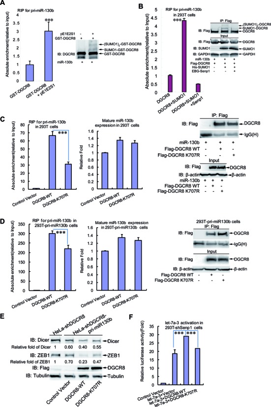
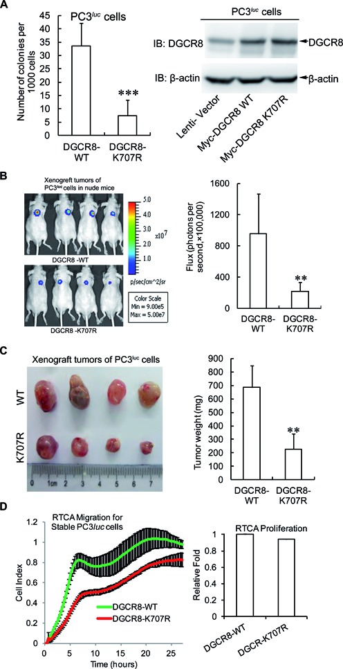
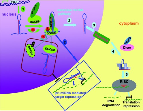
DGCR8 (DiGeorge syndrome critical region gene 8) is essential for primary microRNA (pri-miRNA) processing in the cell nucleus. It specifically combines with Drosha, a nuclear RNase III enzyme, to form the Microprocessor complex (MC) that cleaves pri-miRNA to precursor miRNA (pre-miRNA), which is further processed to mature miRNA by Dicer, a cytoplasmic RNase III enzyme. Increasing evidences suggest that pri-/pre-miRNAs have direct functions in regulation of gene expression, however the underlying mechanism how it is fine-tuned remains unclear. Here we find that DGCR8 is modified by SUMO1 at the major site K(707), which can be promoted by its ERK-activated phosphorylation. SUMOylation of DGCR8 enhances the protein stability by preventing the degradation via the ubiquitin proteasome pathway. More importantly, SUMOylation of DGCR8 does not alter its association with Drosha, the MC activity and miRNA biogenesis, but rather influences its affinity with pri-miRNAs. This altered affinity of DGCR8 with pri-miRNAs seems to control the direct functions of pri-miRNAs in recognition and repression of the target mRNAs, which is evidently linked to the DGCR8 function in regulation of tumorigenesis and cell migration. Collectively, our data suggest a novel mechanism that SUMOylation of DGCR8 controls direct functions of pri-miRNAs in gene silencing.


Figures

Figure 1.
Figure 1.
DGCR8 is modified by SUMO1. (A) DGCR8 is modified mainly by SUMO1. 293T cells were co-transfected with Flag-DGCR8 and three SUMO isoforms along with or without HA-Ubc9. Forty-eight hours after transfection, cells were lysed and pulled down with Ni2+-NTA resin for SUMOylation assay, and SUMOylated modification of DGCR8 was detected with anti-Flag antibody. (B) Endogenous DGCR8 is modified by SUMO1 at multiple sites. His-SUMO with or without Flag-Ubc9 were co-transfected into 293T cells and the SUMOylation assay were conducted with the method of Ni2+-NTA resin. (C) SUMOylation of DGCR8 occurs naturally in cells. PC3 cells of or Senp1−/− MEFs were directly lysed in NEM-RIPA buffer, then immunoprecipitated complexes with anti-DGCR8 or normal IgG were immunoblotted with anti-SUMO1 antibody, and the same membrane was stripped for immunoblotting with anti-DGCR8 antibody. One-tenth of lysates as an input were immunoblotted. (D) SUMOylation of DGCR8 can be removed by Senp1. Flag-DGCR8 with or without His-SUMO1 or EBG-Senp1 plasmids were transfected into 293T cells. The SUMOylation assay with Ni2+-NTA resin was performed. (E and F) SUMOylation of DGCR8 is enhanced by EGF. Flag-DGCR8 and His-SUMO1 were transfected into 293T cells. (E) Twenty-four hours after transfection cells were incubated in serum-free medium for 24 h, then EGF (200 μg/ml) was added for 5 min. (F) Forty-eight hours after transfection cells were treated with U0126 (10 μM) for 1 h. Half of the cells were used for the SUMOylation assay with Ni2+-NTA resin (upper panels), while others lysed with RIPA buffer were for IP with anti-Flag antibody and followed by immunoblotting with pT-P/pS-P antibody (low panels). One-tenth of lysates in RIPA buffer as input were detected with anti-pERK, total ERK antibodies (middle panels). The relative fold of SUMO1-DGCR8 was analyzed by ImageJ (V1.45).
Figure 2.
Figure 2.
(A and B) K707 is a major SUMO-site of DGCR8. 293T cells co-tranfected with DGCR8 WT or mutants K426R, K640R, K707R, K181R, K456R, K510R, K650R or K707/640R with or without His-SUMO1 were lysed for the SUMOylation assay with Ni2+-NTA resin. (C) SUMOylation at K707 of DGCR8 confirmed by IP method. 293T cells transfected with Flag-DGCR8-WT or -K707R with or without GFP-SUMO1 were lysed in NEM-RIPA buffer for immunoprecipitation with anti-Flag antibody and then immunoblotting with anti-GFP antibody. The same membrane was detected with anti-DGCR8 antibody after stripping. One-tenth of lysates as input were analyzed with indicated antibodies. (D) SUMOylation of DGCR8 at K707 is verified by in vitro Escherichia coli-based SUMOylation reconstitution assay. E. coli BL21 cells transformed with GST-DGCR8-WT or -K707R and pE1E2SUMO1 were cultured at 16°C in the presence of 0.2 mM IPTG. GST-DGCR8 proteins were purified and analyzed by immunoblotting with anti-SUMO1 (upper panel) or anti-DGCR8 (low panel) antibody.
Figure 3.
Figure 3.
SUMOylation of DGCR8 enhances its protein stability. (A) Either endogenous or ectopic DGCR8 is more stabilized in Senp1 knockdown 293T or HeLa cells. Indicated 293T and HeLa-shControl or -shSenp1 cells transfected with or without Flag-DGCR8 were lysed in SDS-lysis buffer and then subjected to western blotting for detection of the protein level of endogenous DGCR8 or Flag-DGCR8. (B) Half-life of DGCR8-K707R is shorter than that of DGCR8-WT. HeLa-shDGCR8 cells were transfected with Flag-DGCR8-WT or K707R. Twenty-four hours after transfection cells were treated with 100 μg/ml cycloheximide (CHX) as indicated time. Cells were lysed for immunoblotting analysis. Quantification was analyzed by ImageJ (V1.45) and the Flag-DGCR8 bands were normalized with the Tubulin bands. (C) DGCR8 degrades mainly through the proteasome pathway. HeLa cells were transfected with Flag-DGCR8, 36 h after transfection cells were treated with 40 μM MG132 or 100 μM chloroquine for 6 h and then harvested for immunoblotting analysis. (D) The ubiquitination level of DGCR8-K707R is higher than that of DGCR8-WT. Flag-DGCR8-WT or -K707R with or without Myc-ub were transfected into HeLa cells, 48 h after transfection cells were lysed in RIPA buffer and immunoprecipited with anti-Flag antibody, followed by immunoblotting with anti-Myc antibody. One-tenth of lysates as input were analyzed with indicated antibodies. The relative ratios of all ubiquitin bands of lane 4 and lane 5 in IP was analyzed by ImageJ (V1.45). (E) The protein level of DGCR8 is paralleled with its SUMOylation levels. Flag-DGCR8, His-SUMO1 and Flag-Ubc9 were co-transfected into HeLa cells, 24 h after transfection cells were incubated in serum-free medium for 24 h, then 10 μM U0126 was added for 1 h and followed with treatment of 200 μg/ml EGF for 5 min before harvesting. Cells were lysed for the SUMOylation assay with Ni2+-NTA resin. (F) DGCR8 can be synergistically stabilized by phosphylation and SUMOylation. Flag-DGCR8 along with or without His-SUMO1 and HA-Erk were transfected into HeLa cells, 48 h later, cells were harvested by SDS buffer.
Figure 4.
Figure 4.
SUMOylation of DGCR8 has little effect on the microprocessor activity and miRNA biogenesis. (A) The SUMO-site mutation K707R of DGCR8 does not alter its interaction with Drosha. Lysates from 293T or HeLa cells transfected with Flag-DGCR8-WT or -K707R were used for immunoprecipitation with anti-Flag antibody and then immunoblotting with anti-Drosha antibody. One-tenth of lysates as input were analyzed by immunoblotting. (B) There is no significant difference in the expression level of mature miRNA between low and high levels of DGCR8 SUMOylation. Pri-miR130b and Flag-DGCR8 with or without His-SUMO1 plasmids were transfected into stable cell lines 293T-shControl or -shSenp1. Forty-eight hours after transfection, half of the cells were used for extraction of total RNA, while the others were lysed for the SUMOylation assay with Ni2+-NTA resin. The expression levels of mature miR130b were analyzed by qRT-PCR (left panel) and the SUMOylation levels of DGCR8 were determined by western blotting (right panel). (C and D) SUMOylation of DGCR8 does not affect the microprocessor activity. The microprocessor reporter psiCHECK-pri-miR130b with control vecter, or DGCR8-WT or DGCR8-K707R were transfected into 293T (C) or HeLa-shDGCR8 (D), then 48 h after transfection cells were harvested for the dual-luciferase reporter assay.
Figure 5.
Figure 5.
SUMOylation of DGCR8 increases its affinity with pri-miRNA and silencing effect. (A) SUMOylation of GST-DGCR8 in Escherichia coli enhances its recruiting pri-miRNA. GST-DGCR8 with or without pE1E2S1 expressed in E. coli was purified for RNA pull-down assay. Lysates from 293T cells transfected with pri-miR-130b was added to the same amount of above GST-proteins and GST-beads. After incubation and washing, one-tenth of the combination was subjected to western blotting, while nine-tenth was treated with Trizol for RNA purification and followed by qRT-PCR for pri-miR130b. The SUMO1 modification level of GST-DGCR8 was determined (right panel) and the relative recruitment of pri-miR130b by GST-DGCR8 was normalized with total pri-miR-130b in 293T cells (left panel). (B) Association with pri-miR-130b of DGCR8 is enhanced by its SUMO1 modification. Lysates from 293T cells co-transfected with pri-miR-130b, Flag-DGCR8, His-SUMO1 with or without Senp1 were used for RIP assay with anti-Flag antibody. After incubation and washing, one-tenth of the combination was subjected to western blotting, while 9/10 was treated with Trizol for RNA purification and followed by qRT-PCR for pri-miR130b. The relative recruitment of pri-miR130b by DGCR8 in RIP was normalized with total pri-miR-130b in 293T cells (left panel), and the SUMOylation level and IP efficiency were assessed by western blotting (right panel). (C and D) K707-SUMOylation of DGCR8 influences its binding with pri-miRNA. Lysates from (C) 293T cells co-transfected with pri-miR-130b and Flag-DGCR8-WT or -K707R, or from (D) 293T-pri-miR130b cells transfected with Flag-DGCR8-WT or -K707R, were immunoprecipitated with anti-Flag antibody and then treated with Trizol followed by qRT-PCR for pri-miR130b. The relative recruitment of pri-miR130b by DGCR8 was calculated by normalizing pri-miR130b from the immunoprecipition to the Input group (left panel). The expression level of mature miR130b was analyzed by qRT-PCR (middle panel) and the immunoprecipitation was assessed by western blotting (right panel). (E) The efficiency of pri-miRNA direct function in silencing target gene mediated by DGCR8 is dependent on its K707-SUMOylation. The stable cell line HeLa-shDGCR8-pri-miR130b was transfected with control vector, or DGCR8-WT or DGCR8-K707R for re-expression. HeLa-shDGCR8 cells were used as a control. Forty-eight hours after transfection, cells were lysed in SDS-lysis buffer for detection of Dicer and ZEB1 by western blotting. Quantification was analyzed by ImageJ (V1.45) and the Dicer or ZEB1 bands were normalized with the Tubulin bands. (F) K707-SUMOylation of DGCR8 regulates pri-let-7a-3 activation. The reporter construct psiCHECK-4LCS*let-7a3 and Flag-DGCR8-WT or -K707R with or without pri-let-7a-3 were transfected into 293T-shsenp1 cells, then 48 h after transfection cells were harvested for the dual-luciferase reporter assay.
Figure 6.
Figure 6.
SUMOylation at K707 of DGCR8 promotes tumorigenesis and tumor cell migration. (A) DGCR8-K707R reduces the colony formation of PC3luc cells. Each of 1.5 × 10³ cells stably expressing DGCR8-WT or DGCR8-K707R were seeded in 2 ml of medium containing 1% FBS with 0.35% agar and layered onto the base with 0.6% agar. The colonies were stained with 0.005% crystal violet at day 21, and then photographs were taken and the number of colonies was scored by ImageJ V1.45 (NIH, USA). Three independent experiments were performed in triplicate. (B and C) DGCR8-K707R suppresses tumor growth in nude mice. (B) Backs of 6-week-old nude mice were subcutaneously injected with 2.5 × 10⁶ PC3luc cells stably expressing DGCR8-WT or -K707R. After injection for 14 days, tumor was assessed by bioluminescent imaging with a Xenogen IVIS imaging system and the tumor bioluminescent flux was quantified. (C) All mice were sacrificed at 30 days and tumors were dissected, photographed and weighted. (D) SUMOylation of DGCR8 affects tumor cell migration. For RTCA-Migration assay, 2 × 10⁴ of PC3luc cells stably expressing DGCR8-WT or -K707R were seeded into the upper chambers of the CIM-plate, and normal growth medium containing 10% FBS was added into the lower chamber. The kinetic cell indexes of their migration were recorded every 15 min (Left panel). For RTCA-proliferation assay, 2 × 10³ of cells were seeded into the E-Plate16. The real-time recording of proliferation was carried out on the RTCA-DP instrument (Roche) and monitored every 1 h for 3 days. The relative slope value of cell proliferation was calculated according to the instrument's instruction (right panel).
Figure 7.
Figure 7.
A model for DGCR8 SUMOylation controlling of pri-miRNA direct function. Generally, miRNAs are firstly transcribed by RNA ploymerase II termed as pri-miRNAs, then it is cleaved by Drosha-DGCR8 Microprocessor complex to pre-miRNAs in nucleus. Pre-miRNNAs are transported by Exportin5 to cytoplasm, where the second cleavages are undertaken by Dicer to form a duplex. Finally, the mature miRNA is loaded into the RISC complexes and performs the gene silencing by degradation of the target mRNA or repressing its translation. Apart from as a biogenesis intermediate only, pri-miRNAs exercise direct functions in recognition and repression of the target mRNAs (marked with dark blue Box). DGCR8 SUMOylation at K707 increases its protein stability by preventing the degradation via the ubiquitin proteasome pathway, and its affinity with pri-miRNA thus positively promoting the pri-miRNA direct recognition and repression of the targeted mRNA.

Similar articles

Cited by

References

    1. Han J., Lee Y., Yeom K.H., Kim Y.K., Jin H., Kim V.N. The Drosha-DGCR8 complex in primary microRNA processing. Genes Dev. 2004;18:3016–3027. - PMC - PubMed
    1. Landthaler M., Yalcin A., Tuschl T. The human DiGeorge syndrome critical region gene 8 and Its D. melanogaster homolog are required for miRNA biogenesis. Curr. Biol. 2004;14:2162–2167. - PubMed
    1. Tomari Y., Zamore P.D. MicroRNA biogenesis: drosha can't cut it without a partner. Curr. Biol. 2005;15:R61–R64. - PubMed
    1. Yeom K.H., Lee Y., Han J., Suh M.R., Kim V.N. Characterization of DGCR8/Pasha, the essential cofactor for Drosha in primary miRNA processing. Nucleic Acids Res. 2006;34:4622–4629. - PMC - PubMed
    1. Liu G., Min H., Yue S., Chen C.Z. Pre-miRNA loop nucleotides control the distinct activities of mir-181a-1 and mir-181c in early T cell development. PLoS One. 2008;3:e3592. - PMC - PubMed

LinkOut - more resources