. 2015 Jul 23:4:e06322.
doi: 10.7554/eLife.06322.

Genomic variations of the mevalonate pathway in porokeratosis

Affiliations

Genomic variations of the mevalonate pathway in porokeratosis

Zhenghua Zhang et al. Elife. .

Erratum in

  • Correction: Genomic variations of the mevalonate pathway in porokeratosis.
    Zhang Z, Li C, Wu F, Ma R, Luan J, Yang F, Liu W, Wang L, Zhang S, Liu Y, Gu J, Hua W, Fan M, Peng H, Meng X, Song N, Bi X, Gu C, Zhang Z, Huang Q, Chen L, Xiang L, Xu J, Zheng Z, Jiang Z. Zhang Z, et al. Elife. 2016 Jan 27;5:e14383. doi: 10.7554/eLife.14383. Elife. 2016. PMID: 26816331 Free PMC article. No abstract available.

Abstract

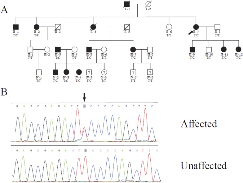
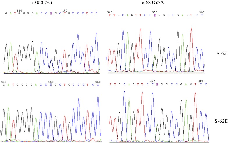
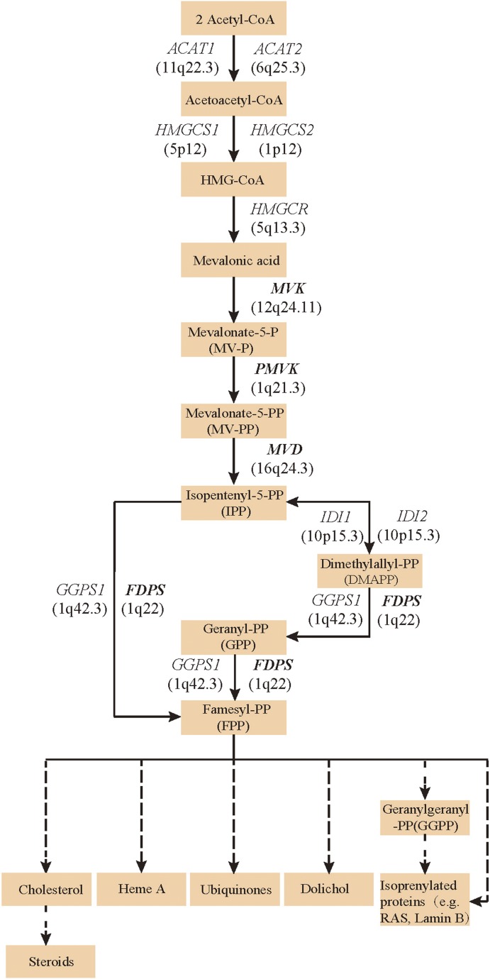
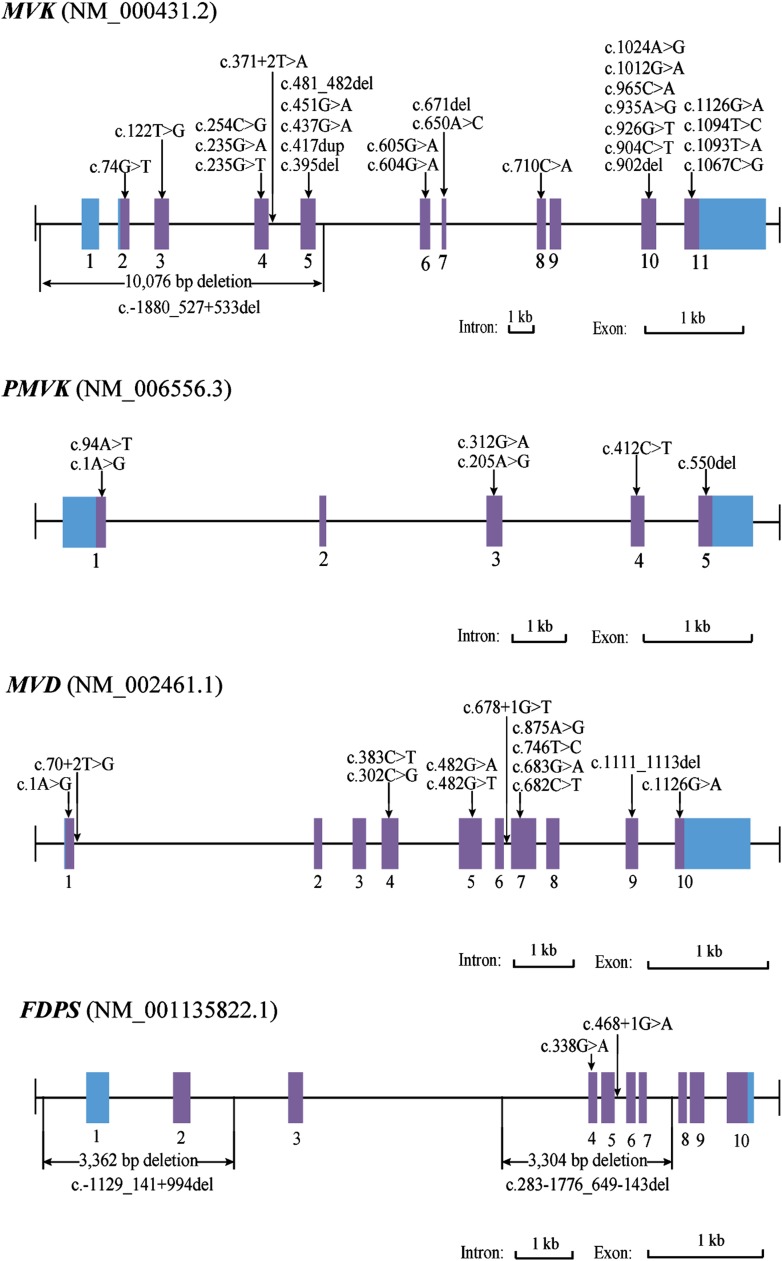
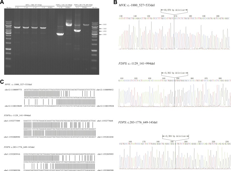
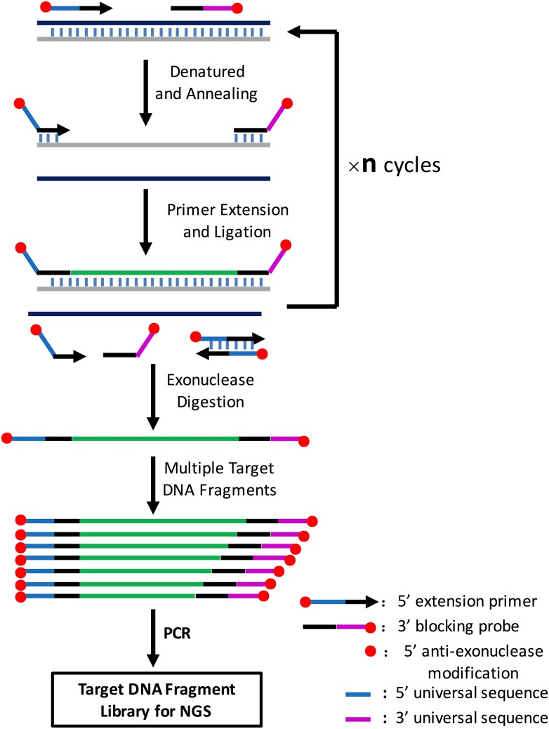
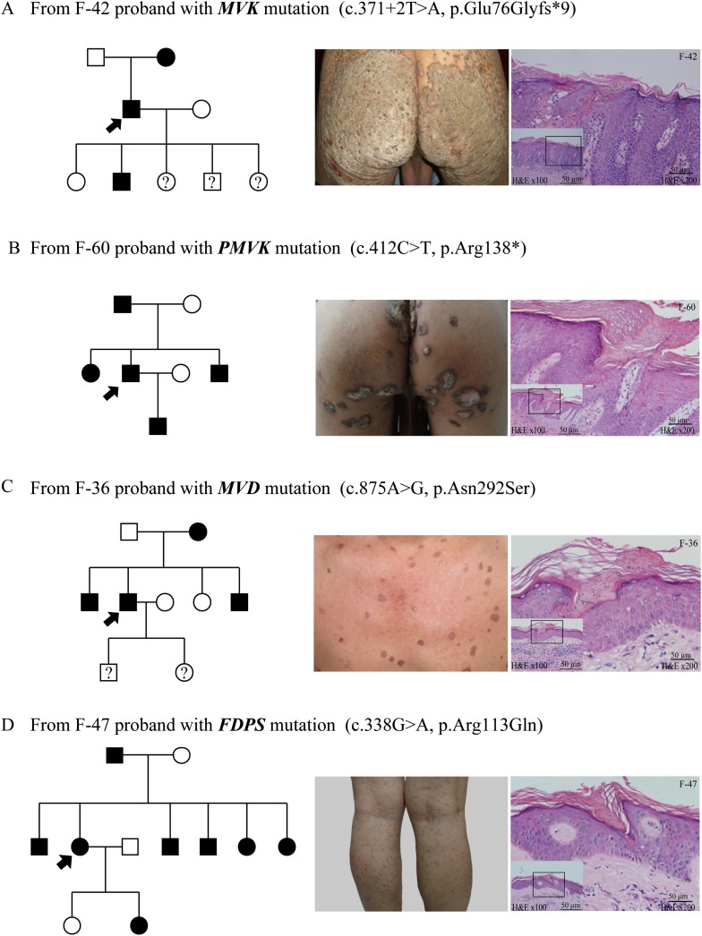
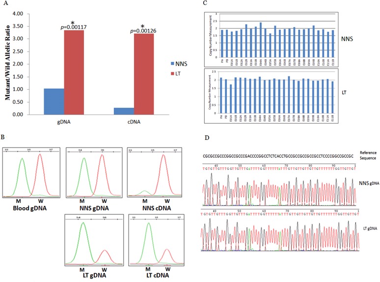
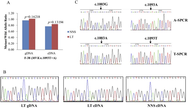
Porokeratosis (PK) is a heterogeneous group of keratinization disorders. No causal genes except MVK have been identified, even though the disease was linked to several genomic loci. Here, we performed massively parallel sequencing and exonic CNV screening of 12 isoprenoid genes in 134 index PK patients (61 familial and 73 sporadic) and identified causal mutations in three novel genes (PMVK, MVD, and FDPS) in addition to MVK in the mevalonate pathway. Allelic expression imbalance (AEI) assays were performed in 13 lesional tissues. At least one mutation in one of the four genes in the mevalonate pathway was found in 60 (98%) familial and 53 (73%) sporadic patients, which suggests that isoprenoid biosynthesis via the mevalonate pathway may play a role in the pathogenesis of PK. Significantly reduced expression of the wild allele was common in lesional tissues due to gene conversion or some other unknown mechanism. A G-to-A RNA editing was observed in one lesional tissue without AEI. In addition, we observed correlations between the mutations in the four mevalonate pathway genes and clinical manifestations in the PK patients, which might support a new and simplified classification of PK under the guidance of genetic testing.

Keywords: genetic testing; human; human biology; medicine; mevalonate pathway; porokeratosis.


Conflict of interest statement

The authors declare that no competing interests exist.

Figures

Figure 1.
Figure 1.. Identification of a MVD mutation in a porokeratosis (PK) family.
(A) c.746T>C (p.Phe249Ser) in MVD displayed 100% co-separation with PK phenotype in this family (Luan et al., 2011). (B) Sanger sequencing chromatograms of proband (II-7, affected) and normal control (II-8, unaffected) at the c.746T>C mutation site indicated by arrow. DOI: http://dx.doi.org/10.7554/eLife.06322.003
Figure 1—figure supplement 1.
Figure 1—figure supplement 1.. Examples of six pedigree charts showing that each mutation displayed 100% co-segregation with the porokeratosis (PK) phenotype in the family.
DOI: http://dx.doi.org/10.7554/eLife.06322.004
Figure 1—figure supplement 2.
Figure 1—figure supplement 2.. Two MVD mutations, c.302C>G (p.Pro101Arg) and c.683 G>A (p.Arg228Gln), for S-62 were located in the trans position because his daughter (S-62-D) carried only one of them.
DOI: http://dx.doi.org/10.7554/eLife.06322.005
Figure 2.
Figure 2.. Isoprenoid biosynthesis via the mevalonate pathway.
12 member genes (ACAT1, ACAT2, HMGCS1, HMGCS2, HMGCR, MVK, PMVK, MVD, IDI1, IDI2, FDPS, GGPS1) were subject to mutation screening. The genomic loci of the 12 member genes are provided in parentheses. The illustration is adapted from the 00900 interactive map of the Kyoto Encyclopedia of genes and genomes (KEGG) (Kanehisa et al., 2012). DOI: http://dx.doi.org/10.7554/eLife.06322.006
Figure 3.
Figure 3.. Mutational spectrum of MVK, PMVK, MVD and FDPS in 113 of the 134 porokeratosis (PK) patients.
DOI: http://dx.doi.org/10.7554/eLife.06322.007
Figure 3—figure supplement 1.
Figure 3—figure supplement 1.. Breakpoint analysis for three large deletion mutations in MVK and FDPS genes.
(A) Agarose electrophoresis analysis of long PCR products from patients with deletion mutations and a normal control. (B) Sequencing chromatograms of long PCR products from deletion mutations. (C) Alignment of the sequences from three deletion mutations with human genome reference GRch38 primary assembly indicates a deletion of 10,076 bp, 3362 bp and 3304 bp, respectively. DOI: http://dx.doi.org/10.7554/eLife.06322.009
Figure 3—figure supplement 2.
Figure 3—figure supplement 2.. Illustration of amplification of multiple target DNA fragments mediated by cycled primer extension and ligation.
In brief, for each target region, an extension primer and a block probe are designed. The extension primer has a 5′ exonuclease-resistance modification and the block probe has a 5′ phosphorate and 3′exonuclease-resistance modification. These two oligos are mixed with genomic DNA, heat-denatured, and then annealed to the same strand of the target DNA fragment. The primer extends and stops until it meets the block probe, and the extension product is then ligated with the block probe by a thermal stable ligase. The above procedure can be repeated n times on the PCR machine using a two-step PCR cycling program. The extension and ligation product is then purified by an exonuclase mixture digestion to remove any DNA fragment with no exonuclease-resistance modification at both ends such as remained primers or probes, primer dimers and genomic DNA, and then amplified using universal NGS PCR primer pairs. A CPELA reaction can include hundreds of extension primer-blocking probe sets and simultaneously amplify hundreds of target DNA fragments for subsequent massively parallel sequencing. DOI: http://dx.doi.org/10.7554/eLife.06322.010
Figure 3—figure supplement 3.
Figure 3—figure supplement 3.. The principle of CNVplex technology.
DOI: http://dx.doi.org/10.7554/eLife.06322.011
Figure 4.
Figure 4.. Representative clinical phenotypes and histopathology associated with the four genotypes.
From left to right, pedigree charts, clinical phenotypes and the corresponding histopathology photos are shown correspondingly. (A) Family (F)-42 proband with MVK mutation showed giant hyperkeratotic plaque-type porokeratosis ptychotropica. (B) F-60 proband with PMVK mutation showed tumor-like porokeratoma in the genitogluteal region. (C) F-36 proband with MVD mutation showed discrete, red-brown annular keratotic papules or maculopapules on the chest. (D) F-47 proband with FDPS mutation showed multiple, small, superficial, annular papules with thread-like ridges on the legs. All histopathology showed cornoid lamella, a histological hallmark of porokeratosis with vertical columns of parakeratosis overlying an area of hypogranulosis with dyskeratotic cells. DOI: http://dx.doi.org/10.7554/eLife.06322.013
Figure 5.
Figure 5.. Gene conversion of the wild to mutant allele was identified in a buttock lesion from F-31 carrying a MVK mutation of c.395delT.
(A) The mutant/wild allelic ratios in genomic DNA (gDNA) and complementary DNA (cDNA) of lesional tissue (LT) and neighboring normal-appearing skin (NNS). The quantity of the mutant allele was about threefold and 10-fold more than the quantity of the wild allele in gDNA and cDNA, respectively. (B) The chromatograms of single nucleotide extension targeting the c.395delT mutation using the SNaPshot kit for five DNA samples (blood gDNA, NNS gDNA, LT gDNA, NNS cDNA and LT cDNA). The mutant peak was overpresented in both LT gDNA and LT cDNA. (C) No copy number change in genomic DNA of NNS and LT. PA and PB are probes in the promoter region, E01 to E11 designates exon 1 to exon 11, and A or B indicate two different probes in the same exon. (D) The bisulfite sequencing of NNS gDNA and LT gDNA. No methylation was observed for the targeted CpG sites in the promoter region of MVK. DOI: http://dx.doi.org/10.7554/eLife.06322.014
Figure 6.
Figure 6.. G-to-A RNA editing at position 1003 of the wild allele (T) of c.1093T>A in MVK was detected in a left forearm lesion from F-38.
(A) No allelic expression imbalance was observed in lesional tissue (LT) from F-38. (B) A mutation of c.1003G>A was identified in LT cDNA, but not in neighboring normal-appearing skin (NNS) cDNA or LT gDNA. (C) Sequencing the c.1093A and c.1093T allele-specific PCR products indicated the mutant allele(A) of c.1003G>A was in the cis position with the wild allele(T) of c.1093T>A. A-SPCR, c.1093A–specific PCR; T-SPCR, c.1093T-specific PCR. DOI: http://dx.doi.org/10.7554/eLife.06322.015
Figure 7.
Figure 7.. Significantly reduced expression of the wild allele in other nine lesion tissues.
The Student t-test was performed to measure the difference in mutant/wild allelic ratios in genomic DNA (gDNA) or complementary DNA (cDNA) from lesion tissue (LT) and neighboring normal-appearing skin (NNS) for each mutation. The test score (p value) is presented above the LT bars. The asterisk (*) designates a significance level of 1%. F-43a and F-43b indicates the tissue sets of the left forearm and left thigh, respectively, from the same F-43 patient. DOI: http://dx.doi.org/10.7554/eLife.06322.016

References

    1. Abramovits W, Oquendo M. Introduction to autoinflammatory syndromes and diseases. Dermatologic Clinics. 2013;31:363–385. doi: 10.1016/j.det.2013.04.010. - DOI - PubMed
    1. Adzhubei IA, Schmidt S, Peshkin L, Ramensky VE, Gerasimova A, Bork P, Kondrashov AS, Sunyaev SR. A method and server for predicting damaging missense mutations. Nature Methods. 2010;7:248–249. doi: 10.1038/nmeth0410-248. - DOI - PMC - PubMed
    1. Biswas A. Cornoid lamellation revisited: apropos of porokeratosis with emphasis on unusual clinicopathological variants. The American Journal of Dermatopathology. 2015;37:145–155. doi: 10.1097/DAD.0000000000000039. - DOI - PubMed
    1. Chen TJ, Chou YC, Chen CH, Kuo TT, Hong HS. Genital porokeratosis: a series of 10 patients and review of the literature. The British Journal of Dermatology. 2006;155:325–329. doi: 10.1111/j.1365-2133.2006.07295.x. - DOI - PubMed
    1. Cui H, Li L, Wang W, Shen J, Yue Z, Zheng X, Zuo X, Liang B, Gao M, Fan X, Yin X, Shen C, Yang C, Zhang C, Zhang X, Sheng Y, Gao J, Zhu Z, Lin D, Zhang A, Wang Z, Liu S, Sun L, Yang S, Cui Y, Zhang X. Exome sequencing identifies SLC17A9 pathogenic gene in two Chinese pedigrees with disseminated superficial actinic porokeratosis. Journal of Medical Genetics. 2014;51:699–704. doi: 10.1136/jmedgenet-2014-102486. - DOI - PubMed

Publication types

MeSH terms

Substances