. 2015 Jul 13:10:4479-90.
doi: 10.2147/IJN.S84930. eCollection 2015.

Magnetic resonance-guided regional gene delivery strategy using a tumor stroma-permeable nanocarrier for pancreatic cancer

Affiliations

Magnetic resonance-guided regional gene delivery strategy using a tumor stroma-permeable nanocarrier for pancreatic cancer

Qingbing Wang et al. Int J Nanomedicine. .

Abstract

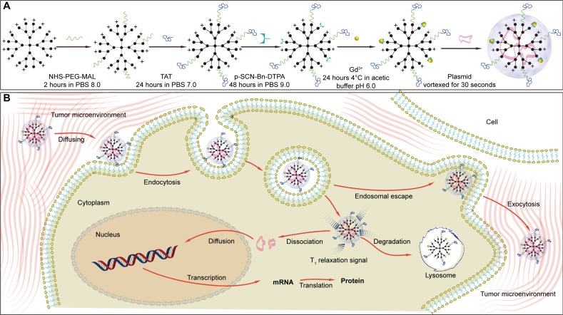
Background: Gene therapy is a very promising technology for treatment of pancreatic ductal adenocarcinoma (PDAC). However, its application has been limited by the abundant stromal response in the tumor microenvironment. The aim of this study was to prepare a dendrimer-based gene-free loading vector with high permeability in the tumor stroma and explore an imaging-guided local gene delivery strategy for PDAC to promote the efficiency of targeted gene delivery.

Methods: The experimental protocol was approved by the animal ethics committee of Zhongshan Hospital, Fudan University. Third-generation dendrigraft poly-L-lysines was selected as the nanocarrier scaffold, which was modified by cell-penetrating peptides and gadolinium (Gd) chelates. DNA plasmids were loaded with these nanocarriers via electrostatic interaction. The cellular uptake and loaded gene expression were examined in MIA PaCa-2 cell lines in vitro. Permeability of the nanoparticles in the tumor stroma and transfected gene distribution in vivo were studied using a magnetic resonance imaging-guided delivery strategy in an orthotopic nude mouse model of PDAC.

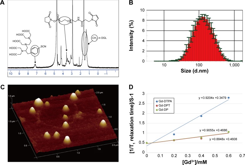
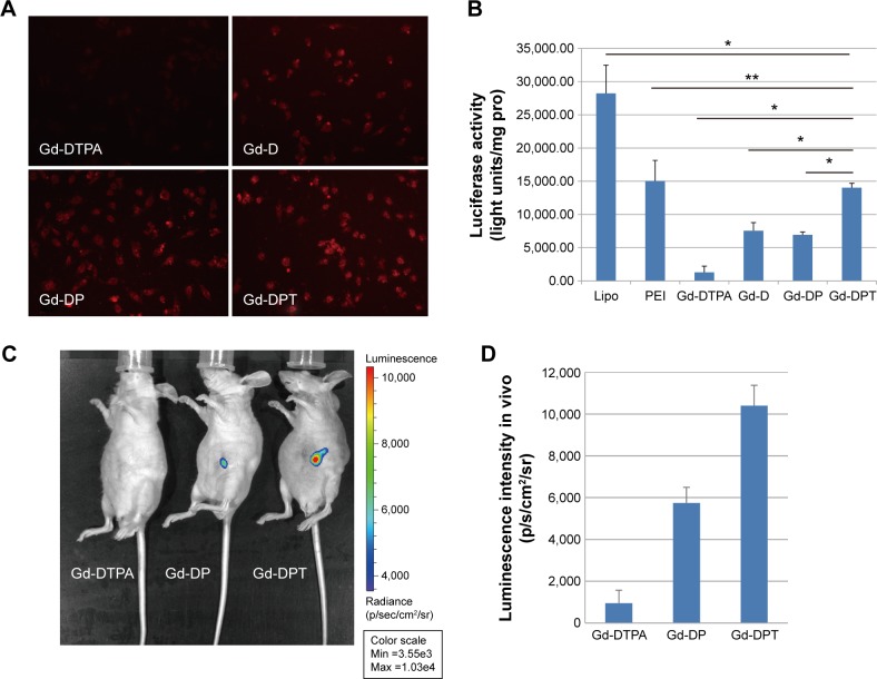
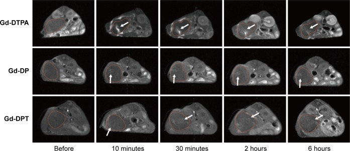
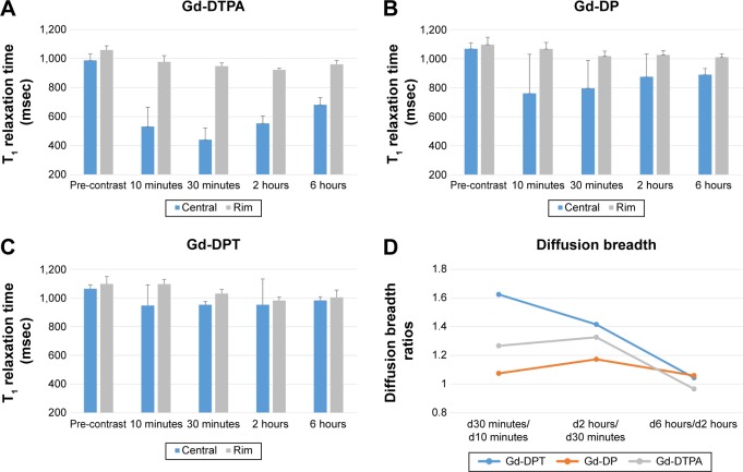
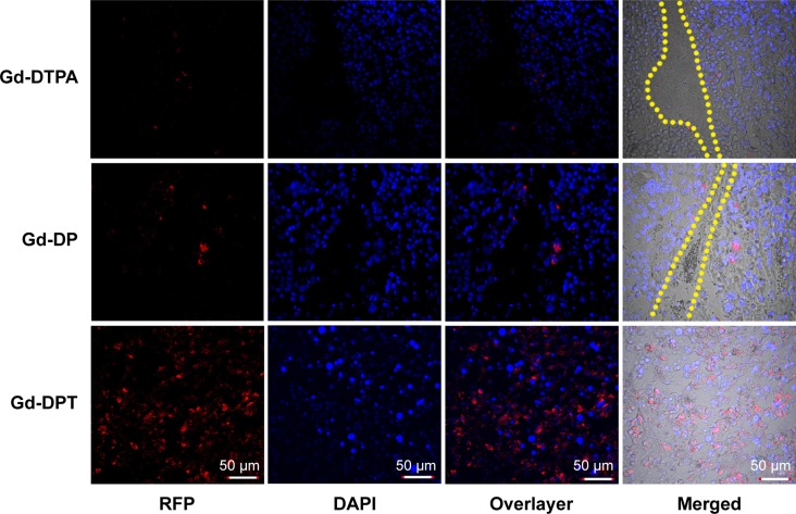
Results: The nanocarriers were synthesized with a dendrigraft poly-L-lysine to polyethylene glycol to DTPA ratio of 1:3.4:8.3 and a mean diameter of 110.9±7.7 nm. The luciferases were strictly expressed in the tumor, and the luminescence intensity in mice treated by Gd-DPT/plasmid luciferase (1.04×10(4)±9.75×10(2) p/s/cm(2)/sr) was significantly (P<0.05) higher than in those treated with Gd-DTPA (9.56×10(2)±6.15×10 p/s/cm(2)/sr) and Gd-DP (5.75×10(3)± 7.45×10(2) p/s/cm(2)/sr). Permeability of the nanoparticles modified by cell-penetrating peptides was superior to that of the unmodified counterpart, demonstrating the improved capability of nanoparticles for diffusion in tumor stroma on magnetic resonance imaging.

Conclusion: This study demonstrated that an image-guided gene delivery system with a stroma-permeable gene vector could be a potential clinically translatable gene therapy strategy for PDAC.

Keywords: cell-penetrating peptides; genetic therapy; interventional; magnetic resonance imaging; molecular imaging; pancreatic cancer.


Figures

Figure 1
Figure 1
Schematic illustration of nanoparticle synthesis and delivery procedure. Notes: (A) Cell-penetrating peptides (TAT) were linked using MAL-PEG-NHS. Paramagnetic Gd3+ ions were chelated with p-SCN-Bn-DTPA conjugated with dendrigraft poly-L-lysine. (B) Nanoparticles were injected intratumorally, and diffused by crossing the stroma, endocytosis, and exocytosis in the tumor microenvironment, all of which were monitored by magnetic resonance imaging. Abbreviations: Gd, gadolinium; MAL, maleimide; NHS, N-hydroxysuccinimide; PEG, polyethylene glycol; PBS, phosphate-buffered saline; p-SCN-Bn-DTPA, 2-(4-isothiocyanatobenzyl)-diethylenetriaminepentaacetic acid.
Figure 2
Figure 2
Nanoparticle characterization. Notes: (A) 1H nuclear magnetic resonance spectra for DTPA-DGL-PEG-TAT. (B) Diameter distribution of Gd-DPT/pRFP. (C) Atomic force microscopy image of Gd-DPT/pRFP. (D) Relaxivity (r1) measurement for Gd-DTPA (3.92 mM−1s−1), Gd-DP/pRFP (0.91 mM−1s−1), and Gd-DPT/pRFP (0.88 mM−1s−1). Abbreviations: Gd, gadolinium; DGL, dendrigraft poly-L-lysine; PEG, polyethylene glycol; pRFP, plasmid red fluorescence protein; DTPA, gadopentetate dimeglumine; DP, DTPA-DGL-PEG; DPT, DTPA-DGL-PEG-TAT.
Figure 3
Figure 3
Gene transfection. Notes: (A) Fluorescent images of RFP expression taken 48 hours after transfection with Gd-DTPA/pRFP (Gd-DTPA), Gd-DGL/pRFP (Gd-D), Gd-DP/pRFP (Gd-DP), and Gd-DPT/pRFP (Gd-DPT). Original magnification, 200×. (B) Luciferase expression 48 hours after administration of Lipofectamine 2000, polyethylenimine, Gd-DTPA, Gd-D, Gd-DP, and Gd-DPT in MIA PaCa-2 cells is plotted as light units per mg protein. *P<0.05, **P>0.05. In vivo distribution (C) and quantitative analysis (D) of transgene loaded by Gd-DTPA/pLuci (Gd-DTPA), Gd-DP/pLuci (Gd-DP), and Gd-DPT/pLuci (Gd-DPT), after intratumoral injection. Abbreviations: Gd, gadolinium; DGL, dendrigraft poly-L-lysine; PEG, polyethylene glycol; Lipo, Lipofectamine; PEI, polyethylenimine; pRFP, plasmid red fluorescence protein; pLuci, plasmid luciferase; Gd-DTPA, gadopentetate dimeglumine; DP, DTPA-DGL-PEG; DPT, DTPA-DGL-PEG-TAT; pro, protein.
Figure 4
Figure 4
Magnetic resonance real-time tracking of the spread of the gene vector in vivo. Before and after injection locally, diffusion of Gd-DTPA/pRFP (Gd-DTPA), Gd-DP/pRFP (Gd-DP), and Gd-DPT/pRFP (Gd-DPT) in the tumor were monitored by T1 weighted imaging (white arrows). For Gd-DTPA, spread in tumor tissue was limited and washout was rapid; for Gd-DP, spread was limited but washout was difficult; and for Gd-DPT, the spread was homogeneous and washout was slow. The tumors are outlined with yellow dot lines on images. Abbreviations: Gd, gadolinium; pRFP, plasmid red fluorescence protein; DGL, dendrigraft poly-L-lysine; PEG, polyethylene glycol; Gd-DTPA, gadopentetate dimeglumine; DP, DTPA-DGL-PEG; DPT, DTPA-DGL-PEG-TAT.
Figure 5
Figure 5
Quantitative imaging analysis. Notes: T1 relaxation time at the center and rim of the tumors was calculated based on T1 software mapping before and after injection of Gd-DTPA/pRFP (A), Gd-DP/pRFP (B), and Gd-DPT/pRFP (C). To reflect the spread patterns after intratumoral injection, the concept of diffusion breadth was defined as the short axis diameter of the enhanced area in the tumor parenchyma. The ratios of diffusion breadths between successive time points were calculated for each group (D). Abbreviations: Gd, gadolinium; pRFP, plasmid red fluorescence protein; DGL, dendrigraft poly-L-lysine; PEG, polyethylene glycol; DTPA, gadopentetate dimeglumine; DP, DTPA-DGL-PEG; DPT, DTPA-DGL-PEG-TAT; d, diffusion breadth.
Figure 6
Figure 6
Confocal luminescence microscopy showing the in vivo distribution and amount of RFP expressed after transfection by naked plasmid or nanoparticles. Upper panel shows Gd-DTPA/pRFP (Gd-DTPA), middle panel shows Gd-DP/pRFP (Gd-DP), and lower panel shows Gd-DPT/pRFP (Gd-DPT). Red indicates RFP, blue indicates DAPI-stained cell nuclei, and yellow lines indicate crevices in tumor tissue. Original magnification 200×. Abbreviations: Gd, gadolinium; DAPI, 4,6-diamidino-2-phenylindole; pRFP, plasmid red fluorescence protein; DGL, dendrigraft poly-L-lysine; PEG, polyethylene glycol; DTPA, gadopentetate dimeglumine; DP, DTPA-DGL-PEG; DPT, DTPA-DGL-PEG-TAT.

References

    1. Caravan P, Farrar CT, Frullano L, Uppal R. Influence of molecular parameters and increasing magnetic field strength on relaxivity of gadolinium- and manganese-based T1 contrast agents. Contrast Media Mol Imaging. 2009;4:89–100. - PMC - PubMed
    1. Siegel R, Naishadham D, Jemal A. Cancer statistics, 2013. CA Cancer J Clin. 2013;63(1):11–30. - PubMed
    1. Jones S, Zhang X, Parsons DW, et al. Core signaling pathways in human pancreatic cancers revealed by global genomic analyses. Science. 2008;321(5897):1801–1806. - PMC - PubMed
    1. Lunardi S, Muschel RJ, Brunner TB. The stromal compartments in pancreatic cancer: are there any therapeutic targets? Cancer Lett. 2014;343(2):147–155. - PubMed
    1. Heinemann V, Reni M, Ychou M, Richel DJ, Macarulla T, Ducreux M. Tumour-stroma interactions in pancreatic ductal adenocarcinoma: rationale and current evidence for new therapeutic strategies. Cancer Treat Rev. 2014;40(1):118–128. - PubMed

Publication types