. 2015 Jun;6(1):163-98.
doi: 10.5598/imafungus.2015.06.01.11. Epub 2015 Jun 11.

The Genera of Fungi - fixing the application of the type species of generic names - G 2: Allantophomopsis, Latorua, Macrodiplodiopsis, Macrohilum, Milospium, Protostegia, Pyricularia, Robillarda, Rotula, Septoriella, Torula, and Wojnowicia

Affiliations

The Genera of Fungi - fixing the application of the type species of generic names - G 2: Allantophomopsis, Latorua, Macrodiplodiopsis, Macrohilum, Milospium, Protostegia, Pyricularia, Robillarda, Rotula, Septoriella, Torula, and Wojnowicia

Pedro W Crous et al. IMA Fungus. 2015 Jun.

Abstract

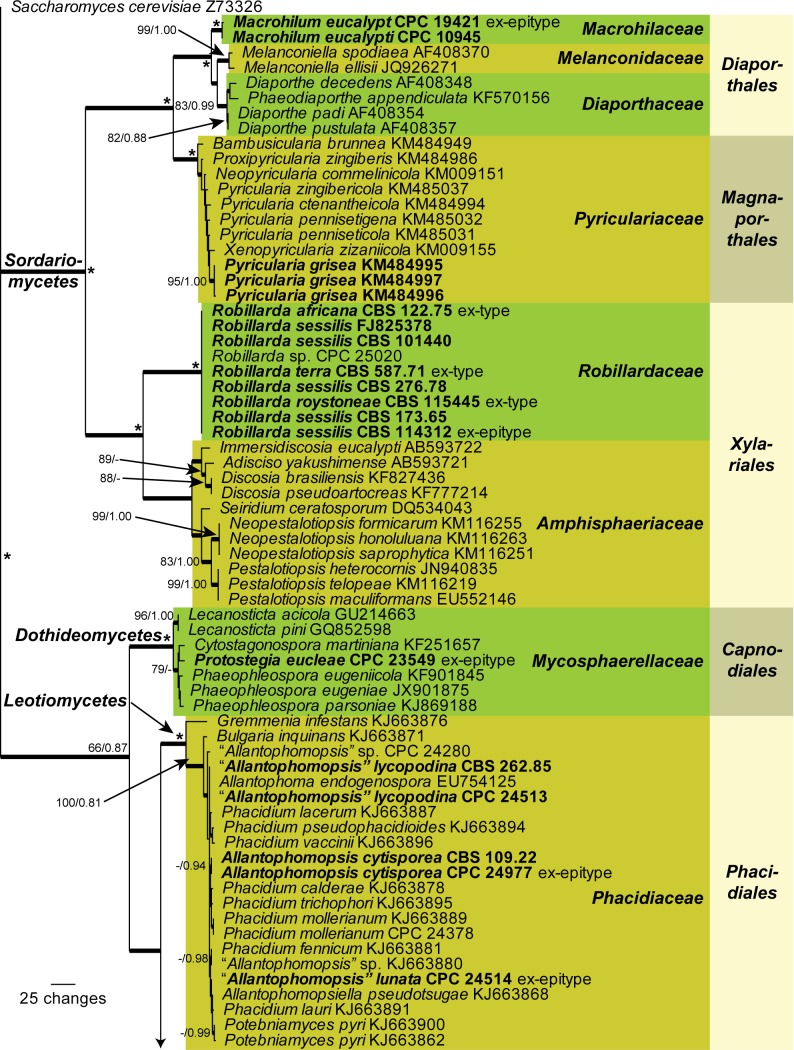
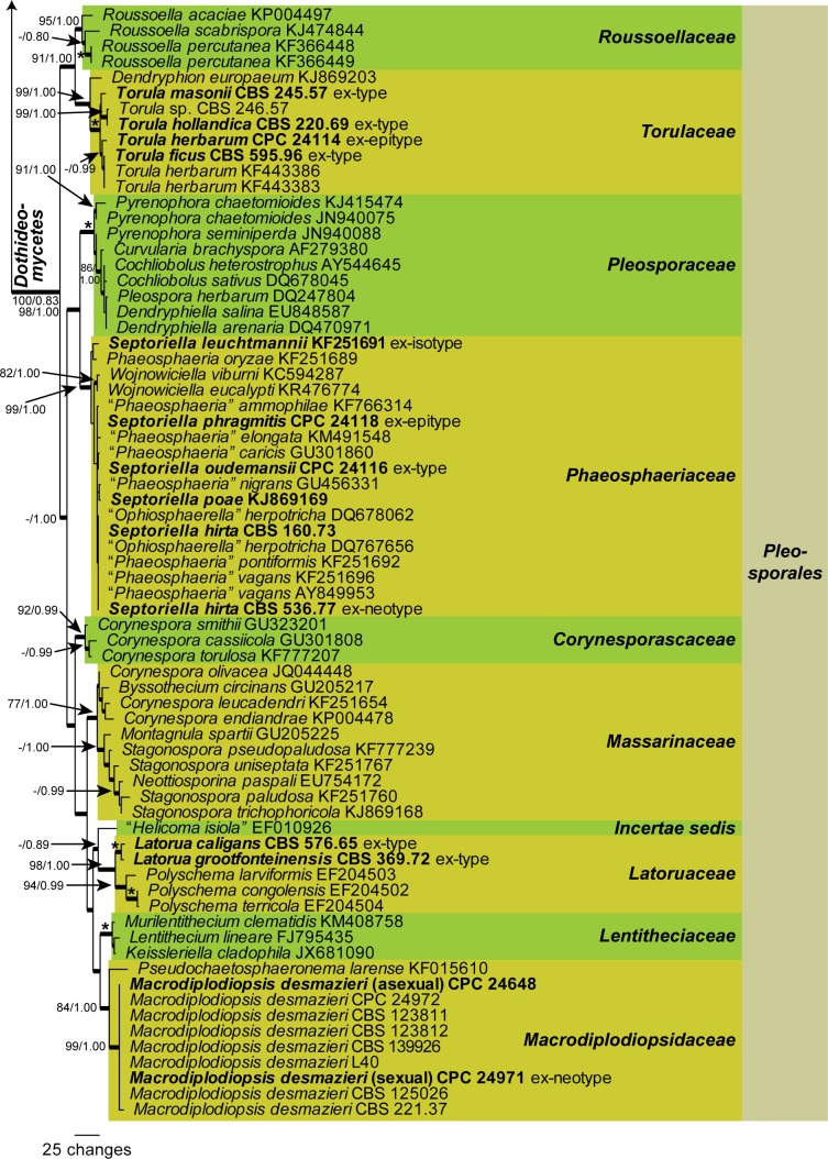
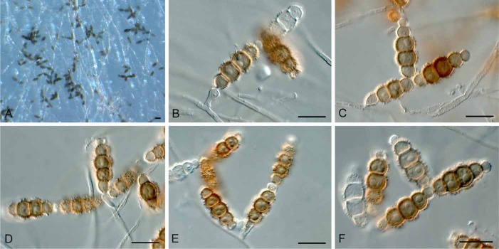
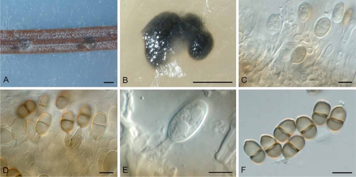
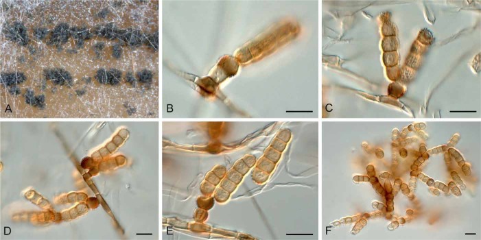
The present paper represents the second contribution in the Genera of Fungi series, linking type species of fungal genera to their morphology and DNA sequence data, and where possible, ecology. This paper focuses on 12 genera of microfungi, 11 of which the type species are neo- or epitypified here: Allantophomopsis (A. cytisporea, Phacidiaceae, Phacidiales, Leotiomycetes), Latorua gen. nov. (Latorua caligans, Latoruaceae, Pleosporales, Dothideomycetes), Macrodiplodiopsis (M. desmazieri, Macrodiplodiopsidaceae, Pleosporales, Dothideomycetes), Macrohilum (M. eucalypti, Macrohilaceae, Diaporthales, Sordariomycetes), Milospium (M. graphideorum, incertae sedis, Pezizomycotina), Protostegia (P. eucleae, Mycosphaerellaceae, Capnodiales, Dothideomycetes), Pyricularia (P. grisea, Pyriculariaceae, Magnaporthales, Sordariomycetes), Robillarda (R. sessilis, Robillardaceae, Xylariales, Sordariomycetes), Rutola (R. graminis, incertae sedis, Pleosporales, Dothideomycetes), Septoriella (S. phragmitis, Phaeosphaeriaceae, Pleosporales, Dothideomycetes), Torula (T. herbarum, Torulaceae, Pleosporales, Dothideomycetes) and Wojnowicia (syn. of Septoriella, S. hirta, Phaeosphaeriaceae, Pleosporales, Dothideomycetes). Novel species include Latorua grootfonteinensis, Robillarda africana, R. roystoneae, R. terrae, Torula ficus, T. hollandica, and T. masonii spp. nov., and three new families: Macrodiplodiopsisceae, Macrohilaceae, and Robillardaceae. Authors interested in contributing accounts of individual genera to larger multi-authored papers to be published in IMA Fungus, should contact the associate editors listed for the major groups of fungi on the List of Protected Generic Names for Fungi (www.generaoffungi.org).

Keywords: DNA Barcodes; ITS; LSU; epitype; fungal systematics; typification; www.GeneraofFungi.org.


Figures

Fig. 1.
Fig. 1.
The first of 1000 equally most parsimonious trees obtained from a heuristic search with 100 random taxon additions of the LSU sequence alignment. Parsimony bootstrap support values >74 % and Bayesian posterior probabilities >0.79 are plotted at the nodes, strict consensus branches from the parsimony analysis are thickened, and the scale bar represents the number of changes. An asterisk (*) denotes a node with 100 % parsimony bootstrap support and 1.00 Bayesian posterior probability. Families and orders are indicated in different colours to the right of the tree and classes at the nodes to the left of the tree. Species treated here for which LSU sequences were available are shown in bold face. The tree was rooted to Saccharomyces cerevisiae (GenBank Z73326).
Fig. 1.
Fig. 1.
The first of 1000 equally most parsimonious trees obtained from a heuristic search with 100 random taxon additions of the LSU sequence alignment. Parsimony bootstrap support values >74 % and Bayesian posterior probabilities >0.79 are plotted at the nodes, strict consensus branches from the parsimony analysis are thickened, and the scale bar represents the number of changes. An asterisk (*) denotes a node with 100 % parsimony bootstrap support and 1.00 Bayesian posterior probability. Families and orders are indicated in different colours to the right of the tree and classes at the nodes to the left of the tree. Species treated here for which LSU sequences were available are shown in bold face. The tree was rooted to Saccharomyces cerevisiae (GenBank Z73326).
Fig. 2.
Fig. 2.
The first of 62 equally most parsimonious trees obtained from a heuristic search with 100 random taxon additions of the ITS sequence alignment. The scale bar shows the number of changes, and bootstrap support values from 1 000 replicates are shown at the nodes. Thickened lines indicate the strict consensus branches and the species treated here are printed in bold face. The species boundaries are delimited with coloured blocks. The tree was rooted to Gremmenia infestans (GenBank accession KM216393).
Fig. 3.
Fig. 3.
The first of two equally most parsimonious trees obtained from a heuristic search with 100 random taxon additions of the combined ITS and HIS sequence alignment. The scale bar shows the number of changes, and bootstrap support values from 1 000 replicates are shown at the nodes. Thickened lines indicate the strict consensus branches and the species treated here are printed in bold face. The species boundaries are delimited with coloured blocks. The tree was rooted to Pestalotiopsis sp. (culture CPC 17179).
Fig. 4.
Fig. 4.
The first of 148 equally most parsimonious trees obtained from a heuristic search with 100 random taxon additions of the combined LSU and ITS sequence alignment. The scale bar shows the number of changes, and bootstrap support values from 1 000 replicates are shown at the nodes (Parsimony bootstrap support / Distance bootstrap support). An asterisk (*) identifies those branches with both 100 % parsimony and distance bootstrap support. Thickened lines indicate the strict consensus branches and the species treated here are printed in bold face. The genus boundaries are delimited with coloured blocks and genus names are printed to the right of the tree in bold face – genus names with inverted commas refer to those genera which do have the generic type species included in the clade whereas those without inverted commas have their generic type species included. The tree was rooted to Didymella exigua (culture CBS 183.55).
Fig. 5.
Fig. 5.
Allantophomopsis cytisporea (CPC 24977). A. Conidioma on PNA. B. Conidiomata forming on OA. C–E. Conidiogenous cells. F. Conidia. Bars: A, B = 600 μm, all others = 10 μm.
Fig. 6.
Fig. 6.
Allantophomopsis lunata (CBS 137781). A, B. Conidiomata on PNA. C. Conidiomata forming on PDA. D. Conidia. Bars: A–C = 250 μm, C = 10 μm.
Fig. 7.
Fig. 7.
Allantophomopsis lycopodina (CBS 262.85). A. Conidioma on PNA. B–D. Conidiogenous cells. E. Conidia (arrows denote mucoid caps). Bars: A = 600 μm, all others = 10 μm.
Fig. 8.
Fig. 8.
Latorua caligans (CBS 576.65). A. Sporulation on SNA. B–F. Conidiogenous cells giving rise to conidial chains. Bars = 10 μm.
Fig. 9.
Fig. 9.
Latorua grootfonteinensis (CBS 369.72). A. Sporulation on SNA. B–G. Conidiogenous cells giving rise to conidial chains. Bars = 10 μm.
Fig. 10.
Fig. 10.
Macrodiplodiopsis desmazieri (CPC 24971). A. Conidioma on host tissue. B–E. Conidiogenous cells. F, G. Conidia (arrows denote mucoid sheaths). H. Asci. I. Ascospores (arrows denote mucoid sheaths). J. Colony on PDA. K. Conidiogenous cells in vitro. L, M. Conidia in vitro (arrows denote mucoid sheaths). Bars: A = 600 μm, J = 10 mm, all others = 10 μm.
Fig. 11.
Fig. 11.
Macrohilum eucalypti (CPC 19421). A. Conidiomata on PNA. B. Conidiomata on OA. C–E. Conidiogenous cells. F. Conidia. Bars: A = 200 μm, B = 300 μm, all others = 10 μm.
Fig. 12.
Fig. 12.
Milospium graphideorum (CBS H-22271). A, B. Dark brown, lichenicolous colonies. C, D. Conidiogenous cells giving rise to lobed conidia. Bars: A, B = 5 mm, C, D = 10 μm.
Fig. 13.
Fig. 13.
Protostegia eucleae (CPC 23549). A. Leaf symptoms. B. Close-up of conidiomata in vivo. C. Vertical section through conidioma. D–F. Conidia. G. Colony on MEA. H. Conidiogenous cells. I. Conidia in vitro. Bars: B = 250 μm, C = 60 μm, G = 5 mm, all others = 10 μm.
Fig. 14.
Fig. 14.
Pyricularia grisea (US0043). A–C. Conidiogenous cells giving rise to conidia. D. Conidia. Bars = 10 μm.
Fig. 15.
Fig. 15.
Robillarda africana (CBS 122.75). A. Conidioma on OA. B, C. Conidiogenous cells. D. Conidia. Bars: A = 200 μm, all others = 10 μm.
Fig. 16.
Fig. 16.
Robillarda roystoneae (CBS 115445). A. Conidiomata on PNA. B, C. Conidiogenous cells. D. Conidia. Bars: A = 200 μm, all others = 10 μm.
Fig. 17.
Fig. 17.
Robillarda sessilis (CBS 114312). A. Conidiomata on PNA. B–E. Conidiogenous cells. F. Conidia. Bars: A = 200 μm, all others = 10 μm.
Fig. 18.
Fig. 18.
Robillarda terrae (CBS 587.71). A. Colony sporulating on OA. B–F. Sections through conidiomata showing wall structure and conidiogenous cells. G–J. Conidia. Bars: A–C = 200 μm, D = 100 μm, all others = 10 μm.
Fig. 19.
Fig. 19.
Rutola graminis (L0054599). A. Colony in vivo. B–G. Conidiogenous cells and conidia. Bars = 10 μm.
Fig. 20.
Fig. 20.
Septoriella phragmitis (CPC 24118). A. Conidiomata in vivo. B. Conidial cirrhus. C, D. Conidiogenous cells. E. Conidia (note mucoid caps). F. Conidia. Bars: A = 300 μm, B = 350 μm, all others = 10 μm.
Fig. 21.
Fig. 21.
Septoriella hirta (CBS 536.77). A, B. Colony on OA (surface and reverse). C. Close-up of conidiomata. D. Conidioma. E, F. Section through conidiomata. G. Conidiogenous cells. H. Developing conidia. I. Mature conidia. Bars: D, E = 100 μm, F = 50 μm, all others = 10 μm.
Fig. 22.
Fig. 22.
Torula ficus (CBS 595.96). A–E. Conidiogenous cells giving rise to conidia. F. Conidia. Bars = 10 μm.
Fig. 23.
Fig. 23.
Torula herbarum (L0118919). A. Colony in vivo. B–D. Conidia. Bars = 10 μm.
Fig. 24.
Fig. 24.
Torula herbarum (CPC 24114). A–D. Conidiogenous cells and conidia. Bars = 10 μm.
Fig. 25.
Fig. 25.
Torula hollandica (CBS 220.69). A. Colony sporulating on OA. B–F. Conidiogenous cells and conidia. Bars = 10 μm.
Fig. 26.
Fig. 26.
Torula masonii (CBS 245.57). A. Colony sporulating on OA. B–H. Conidiogenous cells and conidia. Bars = 10 μm.
Fig. 27.
Fig. 27.
Torula monilis (L0118923). A. Colony in vivo. B–G. Conidiogenous cells and conidia. Bars: B–G = 10 μm.

Similar articles

Cited by

  • Foliar pathogens of eucalypts.
    Crous PW, Wingfield MJ, Cheewangkoon R, Carnegie AJ, Burgess TI, Summerell BA, Edwards J, Taylor PWJ, Groenewald JZ. Crous PW, et al. Stud Mycol. 2019 Aug 8;94:125-298. doi: 10.1016/j.simyco.2019.08.001. eCollection 2019 Sep. Stud Mycol. 2019. PMID: 31636729 Free PMC article.
  • Fungi Present in Antarctic Deep-Sea Sediments Assessed Using DNA Metabarcoding.
    Ogaki MB, Pinto OHB, Vieira R, Neto AA, Convey P, Carvalho-Silva M, Rosa CA, Câmara PEAS, Rosa LH. Ogaki MB, et al. Microb Ecol. 2021 Jul;82(1):157-164. doi: 10.1007/s00248-020-01658-8. Epub 2021 Jan 6. Microb Ecol. 2021. PMID: 33404819
  • Fungal diversity notes 1512-1610: taxonomic and phylogenetic contributions on genera and species of fungal taxa.
    Jayawardena RS, Hyde KD, Wang S, Sun YR, Suwannarach N, Sysouphanthong P, Abdel-Wahab MA, Abdel-Aziz FA, Abeywickrama PD, Abreu VP, Armand A, Aptroot A, Bao DF, Begerow D, Bellanger JM, Bezerra JDP, Bundhun D, Calabon MS, Cao T, Cantillo T, Carvalho JLVR, Chaiwan N, Chen CC, Courtecuisse R, Cui BK, Damm U, Denchev CM, Denchev TT, Deng CY, Devadatha B, de Silva NI, Dos Santos LA, Dubey NK, Dumez S, Ferdinandez HS, Firmino AL, Gafforov Y, Gajanayake AJ, Gomdola D, Gunaseelan S, Shucheng-He, Htet ZH, Kaliyaperumal M, Kemler M, Kezo K, Kularathnage ND, Leonardi M, Li JP, Liao C, Liu S, Loizides M, Luangharn T, Ma J, Madrid H, Mahadevakumar S, Maharachchikumbura SSN, Manamgoda DS, Martín MP, Mekala N, Moreau PA, Mu YH, Pahoua P, Pem D, Pereira OL, Phonrob W, Phukhamsakda C, Raza M, Ren GC, Rinaldi AC, Rossi W, Samarakoon BC, Samarakoon MC, Sarma VV, Senanayake IC, Singh A, Souza MF, Souza-Motta CM, Spielmann AA, Su W, Tang X, Tian X, Thambugala KM, Thongklang N, Tennakoon DS, Wannathes N, Wei D, Welti S, Wijesinghe SN, Yang H, Yang Y, Yuan HS, Zhang H, Zhang J, Balasuriya A, Bhunjun CS, Bulgakov TS, Cai L, Camporesi E, Chomnunti P, Deepika YS, Doilom M, Duan WJ, Han SL, Huanraluek N, J… See abstract for full author list ➔ Jayawardena RS, et al. Fungal Divers. 2022;117(1):1-272. doi: 10.1007/s13225-022-00513-0. Epub 2023 Feb 23. Fungal Divers. 2022. PMID: 36852303 Free PMC article.
  • Neostagonosporellasichuanensis gen. et sp. nov. (Phaeosphaeriaceae, Pleosporales) on Phyllostachysheteroclada (Poaceae) from Sichuan Province, China.
    Yang CL, Xu XL, Wanasinghe DN, Jeewon R, Phookamsak R, Liu YG, Liu LJ, Hyde KD. Yang CL, et al. MycoKeys. 2019 Feb 18;(46):119-150. doi: 10.3897/mycokeys.46.32458. eCollection 2019. MycoKeys. 2019. PMID: 30814907 Free PMC article.
  • Families of Diaporthales based on morphological and phylogenetic evidence.
    Senanayake IC, Crous PW, Groenewald JZ, Maharachchikumbura SSN, Jeewon R, Phillips AJL, Bhat JD, Perera RH, Li QR, Li WJ, Tangthirasunun N, Norphanphoun C, Karunarathna SC, Camporesi E, Manawasighe IS, Al-Sadi AM, Hyde KD. Senanayake IC, et al. Stud Mycol. 2017 Mar;86:217-296. doi: 10.1016/j.simyco.2017.07.003. Epub 2017 Aug 1. Stud Mycol. 2017. PMID: 28947840 Free PMC article.

References

    1. Cannon PF, Damm U, Johnston PR, Weir BS. (2012) Colletotrichum – current status and future directions. Studies in Mycology 73: 181–213. - PMC - PubMed
    1. Carris LM. (1990) Cranberry black rot fungi: Allantophomopsis cytisporea and Allantophomopsis lycopodina. Canadian Journal of Botany 68: 2283–2291.
    1. Cheewangkoon R, Crous PW, Hyde KD, Groenewald JZ, To-anan C. (2008) Species of Mycosphaerella and related anamorphs on Eucalyptus leaves from Thailand. Persoonia 21: 77–91. - PMC - PubMed
    1. Clements FE, Shear CL. (1931) The Genera of Fungi. New York: H.W. Wilson.
    1. Crane JL, Schoknecht JD. (1977) Revision of Torula species. Rutola, a new genus for Torula graminis. Canadian Journal of Botany 55: 3013–3019.

LinkOut - more resources