. 2015 Aug 25;9(8):8012-21.
doi: 10.1021/acsnano.5b01552. Epub 2015 Jul 28.

Spatiotemporal Targeting of a Dual-Ligand Nanoparticle to Cancer Metastasis

Affiliations

Spatiotemporal Targeting of a Dual-Ligand Nanoparticle to Cancer Metastasis

Elizabeth Doolittle et al. ACS Nano. .

Abstract

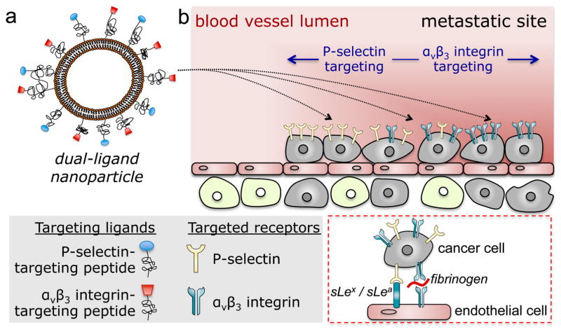
Various targeting strategies and ligands have been employed to direct nanoparticles to tumors that upregulate specific cell-surface molecules. However, tumors display a dynamic, heterogeneous microenvironment, which undergoes spatiotemporal changes including the expression of targetable cell-surface biomarkers. Here, we investigated a dual-ligand nanoparticle to effectively target two receptors overexpressed in aggressive tumors. By using two different chemical specificities, the dual-ligand strategy considered the spatiotemporal alterations in the expression patterns of the receptors in cancer sites. As a case study, we used two mouse models of metastasis of triple-negative breast cancer using the MDA-MB-231 and 4T1 cells. The dual-ligand system utilized two peptides targeting P-selectin and αvβ3 integrin, which are functionally linked to different stages of the development of metastatic disease at a distal site. Using in vivo multimodal imaging and post mortem histological analyses, this study shows that the dual-ligand nanoparticle effectively targeted metastatic disease that was otherwise missed by single-ligand strategies. The dual-ligand nanoparticle was capable of capturing different metastatic sites within the same animal that overexpressed either receptor or both of them. Furthermore, the highly efficient targeting resulted in 22% of the injected dual-ligand nanoparticles being deposited in early-stage metastases within 2 h after injection.

Keywords: cancer metastasis; dual-ligand nanoparticle; triple-negative breast cancer; vascular targeting.


Figures

Figure 1
Figure 1
Illustration of (a) the dual-ligand nanoparticle, and (b) targeting of the nanoparticles to metastatic sites using vascular targeting and a dual-ligand strategy. Inset: Interactions of circulating tumor cells and vascular bed.
Figure 2
Figure 2. Evaluation of the ability of the dual-ligand nanoparticles to target metastasis in vivo in the MDA-MB-231 mouse model
(a) The synopsis shows the timeline and schedule of the in vivo imaging studies. After 25 days from systemic injection of MDA-MB-231 cells via the tail vein, bioluminescence imaging (BLI) showed the development of metastasis in the lungs. Each metastatic site was numbered, which is indicated on the BLI images. (b) Representative fluorescence molecular tomography (FMT) images of the mouse with metastatic spots 4 and 5. FMT imaging was performed 3 h after injection of a cocktail of RGD-NP, PSN-NP and dual-ligand-NP. (c) Using the different NIR fluorophores on each nanoparticle variant, the fluorescence signal in the thoracic region of the FMT images was quantified for each formulation (n=5 mice). Based on phantom measurements of each formulation using the FMT system, the fluorescence signal was converted to nanoparticle concentration (mean±s.d.; y-axis is in logarithmic scale). (d) The total number of nanoparticles for PSN-NP, RGD-NP and dual-ligand-NP is shown for each metastatic spot (y-axis is in logarithmic scale).
Figure 3
Figure 3. Evaluation of the ability of the dual-ligand nanoparticles to target metastasis in vivo in the 4T1 mouse model
(a) The different protocols of in vivo imaging and ex vivo and histological analysis are shown. (b) BLI images show a mouse before and after resection of the primary tumor. (c) ROIs indicate the location of the different organs in an FMT image. (d) Representative FMT images of a mouse with 4T1 metastasis are shown 3 h after injection of a cocktail of RGD-NP, PSN-NP and dual-ligand-NP. After thresholding, the fluorescence signal of each formulation was color-coded (green: RGD-NP; red: PSN-NP; blue: dual-ligand-NP). (e) Using a CRi Maestro fluorescence imaging system, ex vivo imaging of lungs indicated the colocalization of the targeted nanoparticles and 4T1 metastatic cells expressing GFP. (f) Using the ex vivo images to confirm the location of metastatic sites in each mouse, the targeting accuracy of the RGD-NP, PSN-NP and dual-ligand-NP was calculated as a percentage of the metastatic sites being successfully captured by each formulation (n=7 mice; * P<0.05 by Student’s t-test).
Figure 4
Figure 4. Spatiotemporal targeting of P-selectin and αvβ3 integrin in vivo in the 4T1 mouse model
(a) Representative FMT images show targeting of nanoparticles to micrometastases 1 h post-injection in a mouse bearing 4T1 breast cancer metastasis. Mice with 4T1 metastasis (n=8 mice) were injected with a cocktail of αvβ3 integrin-targeting nanoparticles (RGD-NP), P-selectin-targeting nanoparticles (PSN-NP) and non-targeted nanoparticles (NT-NP) containing equal number of particles per formulation. (b) Quantification of the fluorescence signal from hot spots in the FMT images is shown for each formulation. Each animal presented 2–4 hot spots (total 22 hot spots). The animals were imaged at three different time points (t=19, 22 and 26 days after tumor inoculation) to capture early-stage and later stages of metastatic disease. (c) Two hot spots from the same animal show the time-course of signal for RGD-NP and PSN-NP. (d) Comparison of the signal for RGD-NP and PSN-NP was performed for each hot spot in the early-stage (t=19 days) and late-stage disease (t=26 days). To consider a signal of a targeted nanoparticle superior than that of the other formulation, it had to exhibit a difference of 100 pmoles of the fluorophore or greater, which corresponded to the considerable difference of 4×10¹¹ nanoparticles (~10% of the injected dose). Only signals above 80 pmoles of fluorescence were included in the analysis, since this was the detection threshold for the ‘non-specific’ signal of the non-targeted formulation.
Figure 5
Figure 5. Targeting early-stage metastasis using the dual-ligand nanoparticle
(a) The timeline and schedule is shown for the in vivo imaging studies and post mortem analyses. (b) Using whole-body planar gamma scintigraphy, representative coronal image shows the accurate targeting of a 99mTc-labeled dual-ligand-NP to early-stage metastases in the lungs of a mouse with 4T1 metastasis 2 h after injection. Following a systemic injection of ~20 μCi of 99mTc-dual-ligand-NP, the animals were imaged using a Gamma Medica X-SPECT system. The animal model was used 9 days after orthotopic tumor inoculation in a mammary fat pad, which was the time point of early onset of lung metastasis. (c) In the end of the in vivo imaging session, the lungs of the animals were perfused, excised and imaged ex vivo using planar gamma scintigraphy, planar fluorescence imaging and 3D cryo-imaging. 3D-cryo-imaging provided an ultra-high-resolution fluorescence volume of the lungs showing the early onset and topology of metastatic disease. (d) Overlaying ex vivo planar fluorescence and scintigraphy imaging of lungs, the colocalization of dual-ligand-NP and 4T1 metastatic cells is shown. (e) The targeting efficacy of dual-ligand-NP was compared in mice bearing 4T1 metastasis and healthy mice (n=4). Using a gamma scintigraphy, both groups of animals were imaged 2 h after systemic injection of ~20 μCi of 99mTc-dual-ligand-NP. The signal intensity in the thoracic region was compared quantitatively between the two groups.

References

    1. Maeda H, Wu J, Sawa T, Matsumura Y, Hori K. Tumor Vascular Permeability and the EPR Effect in Macromolecular Therapeutics: A Review. J Controlled Release. 2000;65:271–284. - PubMed
    1. Huang X, Peng X, Wang Y, Shin DM, El-Sayed MA, Nie S. A Reexamination of Active and Passive Tumor Targeting by Using Rod-Shaped Gold Nanocrystals and Covalently Conjugated Peptide Ligands. ACS Nano. 2010;4:5887–5896. - PMC - PubMed
    1. Gabizon A, Shmeeda H, Horowitz AT, Zalipsky S. Tumor Cell Targeting of Liposome-Entrapped Drugs with Phospholipid-Anchored Folic Acid-PEG Conjugates. Adv Drug Delivery Rev. 2004;56:1177–1192. - PubMed
    1. Park JW, Hong K, Kirpotin DB, Colbern G, Shalaby R, Baselga J, Shao Y, Nielsen UB, Marks JD, Moore D, et al. Anti-HER2 Immunoliposomes: Enhanced Efficacy Attributable to Targeted Delivery. Clin Cancer Res. 2002;8:1172–1181. - PubMed
    1. Saul JM, Annapragada AV, Bellamkonda RV. A Dual-Ligand Approach for Enhancing Targeting Selectivity of Therapeutic Nanocarriers. J Controlled Release. 2006;114:277–287. - PubMed

Publication types

MeSH terms