

Hairless Streaks in Cattle Implicate TSR2 in Early Hair Follicle Formation
- PMID: 26203908
- PMCID: PMC4512707
- DOI: 10.1371/journal.pgen.1005427
Hairless Streaks in Cattle Implicate TSR2 in Early Hair Follicle Formation
Erratum in
-
Correction: Hairless Streaks in Cattle Implicate TSR2 in Early Hair Follicle Formation.PLoS Genet. 2016 May 2;12(5):e1005688. doi: 10.1371/journal.pgen.1005688. eCollection 2016 May. PLoS Genet. 2016. PMID: 27135403 Free PMC article.
Abstract
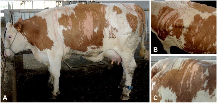
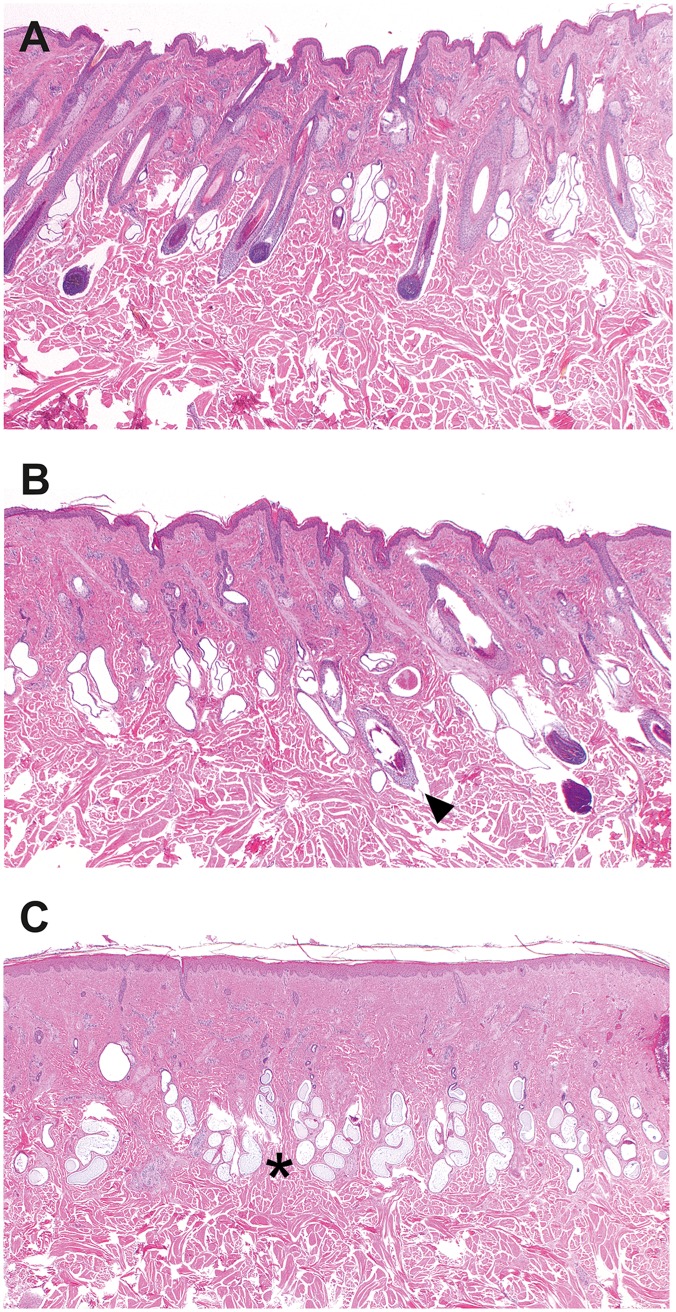
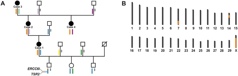
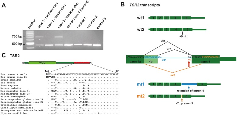
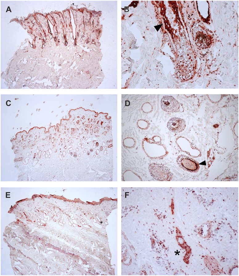
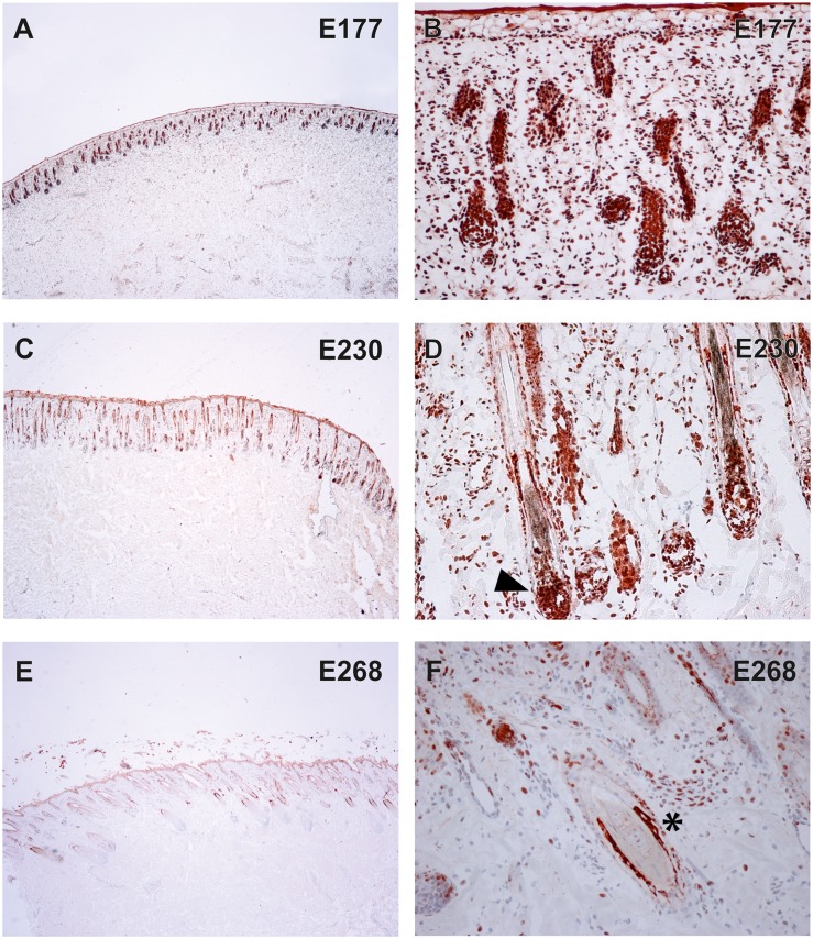
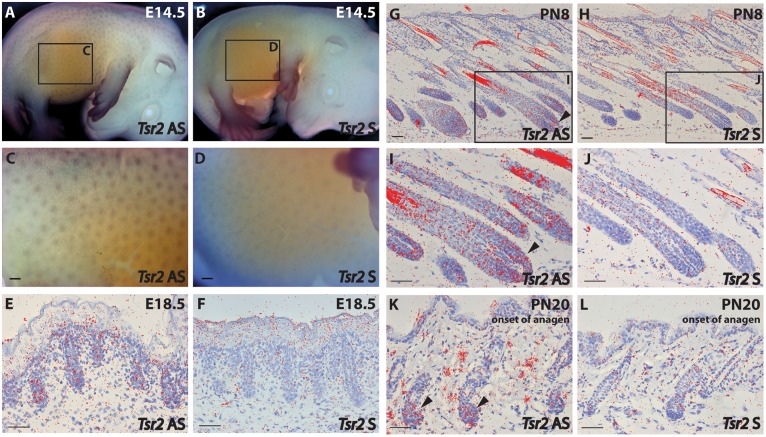
Four related cows showed hairless streaks on various parts of the body with no correlation to the pigmentation pattern. The stripes occurred in a consistent pattern resembling the lines of Blaschko. The non-syndromic hairlessness phenotype observed occurred across three generations of a single family and was compatible with an X-linked mode of inheritance. Linkage analysis and subsequent whole genome sequencing of one affected female identified two perfectly associated non-synonymous sequence variants in the critical interval on bovine chromosome X. Both variants occurred in complete linkage disequilibrium and were absent in more than 3900 controls. An ERCC6L missense mutation was predicted to cause an amino acid substitution of a non-conserved residue. Analysis in mice showed no specific Ercc6l expression pattern related to hair follicle development and therefore ERCC6L was not considered as causative gene. A point mutation at the 5'-splice junction of exon 5 of the TSR2, 20S rRNA accumulation, homolog (S. cerevisiae), gene led to the production of two mutant transcripts, both of which contain a frameshift and generate a premature stop codon predicted to truncate approximately 25% of the protein. Interestingly, in addition to the presence of both physiological TSR2 transcripts, the two mutant transcripts were predominantly detected in the hairless skin of the affected cows. Immunohistochemistry, using an antibody against the N-terminal part of the bovine protein demonstrated the specific expression of the TSR2 protein in the skin and the hair of the affected and the control cows as well as in bovine fetal skin and hair. The RNA hybridization in situ showed that Tsr2 was expressed in pre- and post-natal phases of hair follicle development in mice. Mammalian TSR2 proteins are highly conserved and are known to be broadly expressed, but their precise in vivo functions are poorly understood. Thus, by dissecting a naturally occurring mutation in a domestic animal species, we identified TSR2 as a regulator of hair follicle development.
Conflict of interest statement
The authors have declared that no competing interests exist.
Figures
Similar articles
-
Correction: Hairless Streaks in Cattle Implicate TSR2 in Early Hair Follicle Formation.PLoS Genet. 2016 May 2;12(5):e1005688. doi: 10.1371/journal.pgen.1005688. eCollection 2016 May. PLoS Genet. 2016. PMID: 27135403 Free PMC article.
-
A CHRNB1 frameshift mutation is associated with familial arthrogryposis multiplex congenita in Red dairy cattle.BMC Genomics. 2016 Jun 30;17:479. doi: 10.1186/s12864-016-2832-x. BMC Genomics. 2016. PMID: 27364156 Free PMC article.
-
A mutation in the serum and glucocorticoid-inducible kinase-like kinase (Sgkl) gene is associated with defective hair growth in mice.DNA Res. 2004 Dec 31;11(6):371-9. doi: 10.1093/dnares/11.6.371. DNA Res. 2004. PMID: 15871460
-
The mouse frizzy (fr) and rat 'hairless' (frCR) mutations are natural variants of protease serine S1 family member 8 (Prss8).Exp Dermatol. 2010 Jun;19(6):527-32. doi: 10.1111/j.1600-0625.2009.01054.x. Epub 2010 Feb 25. Exp Dermatol. 2010. PMID: 20201958
-
Independent DSG4 frameshift variants in cats with hair shaft dystrophy.Mol Genet Genomics. 2022 Jan;297(1):147-154. doi: 10.1007/s00438-021-01842-6. Epub 2021 Dec 8. Mol Genet Genomics. 2022. PMID: 34878611 Free PMC article.
Cited by
-
Functional impact of splicing variants in the elaboration of complex traits in cattle.Nat Commun. 2025 Apr 24;16(1):3893. doi: 10.1038/s41467-025-58970-5. Nat Commun. 2025. PMID: 40274775 Free PMC article.
-
Correction: Hairless Streaks in Cattle Implicate TSR2 in Early Hair Follicle Formation.PLoS Genet. 2016 May 2;12(5):e1005688. doi: 10.1371/journal.pgen.1005688. eCollection 2016 May. PLoS Genet. 2016. PMID: 27135403 Free PMC article.
-
An Intronic MBTPS2 Variant Results in a Splicing Defect in Horses with Brindle Coat Texture.G3 (Bethesda). 2016 Sep 8;6(9):2963-70. doi: 10.1534/g3.116.032433. G3 (Bethesda). 2016. PMID: 27449517 Free PMC article.
-
A Large Deletion in the NSDHL Gene in Labrador Retrievers with a Congenital Cornification Disorder.G3 (Bethesda). 2017 Sep 7;7(9):3115-3121. doi: 10.1534/g3.117.1124. G3 (Bethesda). 2017. PMID: 28739597 Free PMC article.
-
A CHRNB1 frameshift mutation is associated with familial arthrogryposis multiplex congenita in Red dairy cattle.BMC Genomics. 2016 Jun 30;17:479. doi: 10.1186/s12864-016-2832-x. BMC Genomics. 2016. PMID: 27364156 Free PMC article.
References
-
- Blaschko A (1901) Die Nervenverteilung in der Haut in ihrer Beziehung zu den Erkrankungen der Haut. Breslau: Beilage zu den Verhandlungen der Deutschen Dermatologischen Gesellschaft VII Congress (German).
-
- Happle R (1985) Lyonization and the lines of Blaschko. Hum Genet 70: 200–206. - PubMed
-
- Happle R (2006) X-chromosome inactivation: role in skin disease expression. Acta Paediatr (S451) 95: 16–23. - PubMed
MeSH terms
Substances
LinkOut - more resources
Full Text Sources
Other Literature Sources
Molecular Biology Databases
