

The Brain Functional State of Music Creation: an fMRI Study of Composers
- PMID: 26203921
- PMCID: PMC4512184
- DOI: 10.1038/srep12277
The Brain Functional State of Music Creation: an fMRI Study of Composers
Abstract
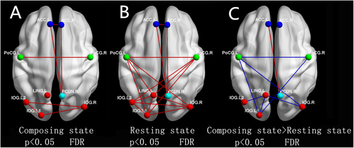
In this study, we used functional magnetic resonance imaging (fMRI) to explore the functional networks in professional composers during the creation of music. We compared the composing state and resting state imagery of 17 composers and found that the functional connectivity of primary networks in the bilateral occipital lobe and bilateral postcentral cortex decreased during the composing period. However, significantly stronger functional connectivity appeared between the anterior cingulate cortex (ACC), the right angular gyrus and the bilateral superior frontal gyrus during composition. These findings indicate that a specific brain state of musical creation is formed when professional composers are composing, in which the integration of the primary visual and motor areas is not necessary. Instead, the neurons of these areas are recruited to enhance the functional connectivity between the ACC and the default mode network (DMN) to plan the integration of musical notes with emotion.
Figures
Similar articles
-
[Altered patterns of functional connectivity of posterior cingulate cortex on resting-state magnetic resonance imaging in children with attention-deficit or hyperactivity disorder].Zhonghua Yi Xue Za Zhi. 2013 Jun 25;93(24):1881-5. Zhonghua Yi Xue Za Zhi. 2013. PMID: 24124739 Chinese.
-
Morphological brain plasticity induced by musical expertise is accompanied by modulation of functional connectivity at rest.Neuroimage. 2014 Apr 15;90:179-88. doi: 10.1016/j.neuroimage.2013.12.065. Epub 2014 Jan 10. Neuroimage. 2014. PMID: 24418502
-
Playing piano in the mind--an fMRI study on music imagery and performance in pianists.Brain Res Cogn Brain Res. 2004 May;19(3):219-28. doi: 10.1016/j.cogbrainres.2003.12.005. Brain Res Cogn Brain Res. 2004. PMID: 15062860
-
Core Concept: Resting-state connectivity.Proc Natl Acad Sci U S A. 2015 Nov 17;112(46):14115-6. doi: 10.1073/pnas.1518785112. Proc Natl Acad Sci U S A. 2015. PMID: 26578753 Free PMC article. Review. No abstract available.
-
Ten Famous Composers of the Romantic Era and Their Causes of Death.Med Princ Pract. 2022;31(1):20-28. doi: 10.1159/000521537. Epub 2021 Dec 17. Med Princ Pract. 2022. PMID: 34923496 Free PMC article. Review.
Cited by
-
Hierarchical dynamics of informational patterns and decision-making.Proc Biol Sci. 2016 Jun 15;283(1832):20160475. doi: 10.1098/rspb.2016.0475. Proc Biol Sci. 2016. PMID: 27252020 Free PMC article.
-
Dimensions of Musical Creativity.Front Neurosci. 2020 Nov 30;14:578932. doi: 10.3389/fnins.2020.578932. eCollection 2020. Front Neurosci. 2020. PMID: 33328852 Free PMC article.
-
The association between liking, learning and creativity in music.Sci Rep. 2024 Aug 16;14(1):19048. doi: 10.1038/s41598-024-70027-z. Sci Rep. 2024. PMID: 39152203 Free PMC article.
-
Individualized goal directed dance rehabilitation in chronic state of severe traumatic brain injury: A case study.Heliyon. 2019 Feb 12;5(2):e01184. doi: 10.1016/j.heliyon.2019.e01184. eCollection 2019 Feb. Heliyon. 2019. PMID: 30805564 Free PMC article.
-
The Multiple-Demand System in the Novelty of Musical Improvisation: Evidence from an MRI Study on Composers.Front Neurosci. 2017 Dec 11;11:695. doi: 10.3389/fnins.2017.00695. eCollection 2017. Front Neurosci. 2017. PMID: 29311776 Free PMC article.
References
-
- Simonton D. K. Origins of genius. Darwinian perspectives on creativity. (Oxford University Press, 1999).
-
- Sternberg R. J. Handbook of creativity. (Cambridge University Press, 1999).
-
- Lau H. C., Rogers R. D., Ramnani N. & Passingham R. E. Willed action and attention to the selection of action. NeuroImage 21, 1407–1415 (2004). - PubMed
-
- Nathaniel-James D. A. & Frith C. D. The role of the dorsolateral prefrontal cortex: evidence from the effects of contextual constraint in a sentence completion task. NeuroImage 16, 1094–1102 (2002). - PubMed
Publication types
MeSH terms
LinkOut - more resources
Full Text Sources
Other Literature Sources
Medical
