. 2015 Jul 23:6:7815.
doi: 10.1038/ncomms8815.

Condensin targets and reduces unwound DNA structures associated with transcription in mitotic chromosome condensation

Affiliations

Condensin targets and reduces unwound DNA structures associated with transcription in mitotic chromosome condensation

Takashi Sutani et al. Nat Commun. .

Abstract

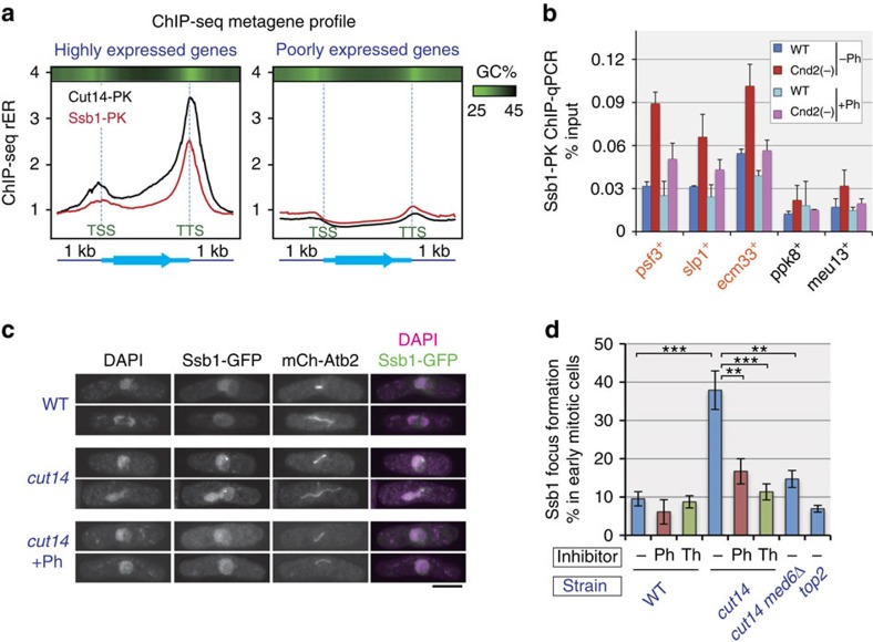
Chromosome condensation is a hallmark of mitosis in eukaryotes and is a prerequisite for faithful segregation of genetic material to daughter cells. Here we show that condensin, which is essential for assembling condensed chromosomes, helps to preclude the detrimental effects of gene transcription on mitotic condensation. ChIP-seq profiling reveals that the fission yeast condensin preferentially binds to active protein-coding genes in a transcription-dependent manner during mitosis. Pharmacological and genetic attenuation of transcription largely rescue bulk chromosome segregation defects observed in condensin mutants. We also demonstrate that condensin is associated with and reduces unwound DNA segments generated by transcription, providing a direct link between an in vitro activity of condensin and its in vivo function. The human condensin isoform condensin I also binds to unwound DNA regions at the transcription start sites of active genes, implying that our findings uncover a fundamental feature of condensin complexes.


Figures

Figure 1
Figure 1. Identification of condensin binding sites in fission yeast mitotic chromosomes.
(a) Binding profile of condensin complex along the chromosome III (chr III) left arm. Cells with an epitope-tagged condensin subunit (Cut14-PK) were arrested at prometaphase by the nda3-KM311 cold-sensitive tubulin allele and analysed by ChIP-seq using anti-PK antibody. The rER (vertical axis) reflects the binding probability at the corresponding genome site (horizontal axis); cen, centromere. (b) Verification of ChIP-seq results by qPCR. DNA isolated by Cut14-PK ChIP was measured by qPCR at 13 selected sites in the genome, including the identified condensin binding sites (Supplementary Fig. 1d). The resulting ChIP efficiency values (represented as % input) showed good correlation with ChIP-seq rERs, verifying the accuracy of ChIP-seq results. r, Pearson's correlation coefficient. (c) Physiological relevance of detected condensin binding. Chromosome binding of epitope-tagged wild-type and temperature-sensitive Cut14 proteins (Cut14-PK and Cut14ts-PK, respectively) was assayed by ChIP-qPCR. Cells were arrested in prometaphase by cultivating at 20 °C, a permissive temperature for cut14-208 but restrictive temperature for the cold-sensitive nda3 mutation. The mutant protein showed reduced binding at all locations tested, including the newly identified Cut14-enriched sites (orange), even at the permissive temperature. Each qPCR site is named after the nearby gene or genomic feature; cnt, central core regions of centromeres 1 and 3. Cut14ts protein is not heat labile and is as stable as the wild-type protein. No tag, untagged wild-type cells as a control. Error bars represent s.d. (n=2, technical replicates in qPCR). (d) Heat map of condensin enrichment for all protein-coding genes from 1 kb upstream of the TSS to 1 kb downstream of the TTS. Gene lengths are scaled to the same size. Genes are ranked from highest to lowest condensin enrichment.
Figure 2
Figure 2. Transcription-dependent condensin binding at RNAP2-driven active genes.
(a) ChIP-seq profiles of condensin (Cut14-PK), RNAP2 (detected by monoclonal antibody 8WG16, recognizing the largest subunit Rpb1) and all RNA polymerases (RNAPs, detected using the PK epitope on the common subunit Rpb5), as well as a poly(A)-selected RNA-seq profile. All profiles are from prometaphase cells. Annotated ORFs (cyan) and other transcripts (non-coding RNA and tRNA, magenta) are shown at the bottom. chr I, chromosome I. (b) Genome-wide correlation plot of Cut14-PK and RNAP2 ChIP-seq results. Purple, green and blue correspond to centromeres (cen), rDNA and tRNA gene loci, respectively. (c) Transcription-dependent condensin binding. Cut14-PK cells in prometaphase were treated with the transcription inhibitor 1,10-phenanthroline (Ph; 60 or 120 μg ml−1) or thiolutin (Th; 20 μg ml−1) for 30 min and analysed by anti-PK or anti-RNAP2 ChIP-qPCR. The translation inhibitor cycloheximide (CHX; 100 μg ml-1) was used as a negative control. Error bars represent s.d. (n=2, technical replicates in qPCR). cnt, central core regions of centromeres 1 and 3.
Figure 3
Figure 3. Suppression of chromosome segregation defect in condensin mutants by transcriptional attenuation.
(a) Wild-type (WT) or temperature-sensitive condensin mutant (cut14-208) cells were arrested at prometaphase, treated with or without a transcription inhibitor (1,10-phenanthroline (Ph; 60 μg ml−1) or thiolutin (Th; 1 μg ml−1)) for 30 min and then released from arrest at the restrictive temperature (34 °C) for 15 min. Cells were fixed, and the nuclear morphology in post-anaphase cells was examined by DAPI staining. The frequency of stretched but unsegregated nuclei in cut14-208 cells was significantly reduced by inhibitor treatment. Error bars represent s.e.m. (n=3, independent experiments). Scale bar, 5 μm. (b) Suppressor mutations in three independent phenotypic revertants of cut3 were revealed by whole-genome sequencing to reside in med6 (Mediator complex subunit 6). X, nonsense mutation; fs, frameshift mutation. (c) Rescue of growth defect in condensin mutants by med6 deletion (med6Δ). Med6 depletion rescued cut3-477 completely at 36 °C and partially suppressed the more severe cut14-208 at 33 °C. (d) Rescue of chromosome segregation defect in condensin mutants by med6Δ. Cells were cultivated at restrictive temperatures (36 °C, 4 h for cut3, cut3 med6Δ; 34 °C, 2 h for cut14, cut14 med6Δ) and then fixed. Nuclear morphology in mitotic cells was revealed by DAPI staining. Scale bar, 5 μm. (e) Genome-wide comparison of RNAP2 binding between WT and med6Δ cells. The amount of RNAP2 bound to each protein-coding gene was calculated based on ChIP-seq and ChIP-qPCR results and plotted. RNAP2 binding was decreased at most genes in med6Δ. Genes monitored by qPCR (Supplementary Fig. 3b) are shown in red.
Figure 4
Figure 4. Presence of ssDNA at condensin binding sites.
(a) Treatment of condensin-bound DNA fragments with nuclease P1, which is specific to ssDNA/single-stranded RNA. DNA fragments purified by Cut14-PK ChIP from prometaphase cells were treated with P1 on beads and then eluted and measured by qPCR (left). P1 sensitivity was specific to condensin-bound fragments, because bulk DNA at the same sites (purified by anti-histone H3 ChIP from prometaphase cells) or cohesin-associated DNA (purified by Rad21-GFP ChIP from asynchronous cells) showed no sensitivity (middle and right, respectively). (b) RNase treatment of condensin-bound DNA fragments. RNase A or RNase H treatment, which digests single-stranded RNA or RNA within DNA:RNA hybrids, respectively, caused no reduction in qPCR measurements, precluding the possibility that the condensin-DNA association is mediated by RNA. Error bars represent s.d. (n=2, technical replicates in qPCR). cnt, central core regions of centromeres 1 and 3.
Figure 5
Figure 5. Opposing roles of transcription and condensin in DNA unwinding.
(a) Metagene ChIP-seq profiles of condensin (Cut14-PK) and the eukaryotic ssDNA-binding factor RPA (Ssb1-PK). Distribution profiles were averaged over highly expressed genes in prometaphase cells (top 10%) as well as poorly expressed genes (bottom 50%). The profile is from 1 kb upstream of the TSS to 1 kb downstream of the TTS, and gene lengths were scaled to the same size. Top bars represent average GC content in a 100-bp window (bright green indicates lower GC content). Ssb1 is co-localized with condensin around the TTS of highly expressed genes. (b) ChIP-qPCR analysis of Ssb1-PK in prometaphase-arrested wild-type and condensin-depleted cells. Condensin depletion increased chromosomal binding of Ssb1 at condensin binding sites (orange), but this effect was attenuated by the transcription inhibitor 1,10-phenanthroline (Ph). Error bars represent s.d. (n=2, technical replicates in qPCR). (c,d) Wild-type (WT), cut14, cut14 med6Δ and top2 cells expressing GFP-tagged Ssb1 and mCherry-tagged Atb2 (encoding α-tubulin) were cultured at 34 °C for 1 h and then fixed. Ph and thiolutin (Th) indicate treatment for the last 30 min with Ph (120 μg ml−1) and Th (2 μg ml−1), respectively. Micrographs of representative mitotic cells (c) and frequency of Ssb1 focus formation in nuclei of early mitotic cells with short spindles (d) are shown. Untreated cut14 cells showed elevated levels of Ssb1 focus formation compared with WT, drug-treated cut14 or cut14 med6Δ cells (**P<0.01 and ***P<0.001; Welch's t-test, one tailed), indicating that transcription promotes and condensin represses the accumulation of unwound DNA segments in mitotic cells. Scale bar, 5 μm. Error bars represent s.e.m. (n=7 independent experiments for cut14, n=4 for cut14+Ph, n=3 for others).
Figure 6
Figure 6. Binding of human condensin I complex to the TSS of active genes transcribed by RNAP2 and RNAP3.
(a,b) ChIP-seq profiles of condensin I (using an antibody against NCAPG), RNAP2 (using 8WG16 monoclonal antibody, a) and RNAP3 (using monoclonal antibody against RPC32 subunit, b) in HeLa cells under the indicated conditions. M, cells arrested in prometaphase by nocodazole; Asy, asynchronous cells. Where indicated, the NCAPG subunit was depleted by siRNA treatment (Supplementary Fig. 8a). The upper boxes show the positions of protein-coding genes, and the lower boxes in b indicate the positions of tRNA genes (those within ORFs are shown in purple; others are shown in orange). The four sets of graphs in a and b show ChIP-seq profiles. The y axes of the ChIP-seq profiles indicate the normalized read intensity. Red indicates regions where enrichment determined by ChIP was statistically significant. (c) Distance of condensin I peaks from TSSs. More than 70% of NCAPG-binding sites are localized within ±5 kb of a TSS. (d) Level of condensin I binding during mitosis correlates with level of transcription during interphase. All RefSeq genes were ranked and divided into four equal groups based on their expression levels (high to low) in asynchronous cells, and averaged condensin I binding profiles around the TSS for each group were calculated. (e) Averaged mitotic NCAPG ChIP-seq profiles around tRNA genes. The profile for tRNA genes associated with RNAP3 in asynchronous cells is shown in purple.
Figure 7
Figure 7. Recognition of ssDNA and expulsion of RNAPs from TSSs by human condensin I.
(a) Appearance of RNAP2 ChIP-seq peaks at TSSs in condensin I−depleted cells. Mitotic ChIP-seq profiles of NCAPG and RNAP2 around TSSs (within ±5 kb) were averaged over all RefSeq genes and plotted. The y axis indicates the normalized read density. Profiles of NCAPG and RNAP2 ChIP-seq as well as RNAP2 ChIP-seq in NCAPG-depleted cells are shown. The inset provides a magnified view of the region ±500 bp from the TSS. (b) ChIP-qPCR of RNAP2 (detected by monoclonal antibody 8WG16) and active RNAP2 (phosphorylated at Ser2 in the C-terminal domain repeats, pS2). The NCAPG condensin I subunit and TTF2 were depleted by siRNA treatment, both individually and in combination. Binding to two gene loci (BRD2 and RPL13) was measured at the TSS, TTS and midpoint of the gene (gene body). NCAPG knockdown promoted binding of RNAP2 at the TSS, but not at the gene body or TTS, indicating that condensin I depletion is insufficient to de-repress mitotic gene expression. (c) Appearance of RNAP3 ChIP-seq peaks at the TSS of tRNA genes in condensin I-depleted cells. Averaged mitotic RNAP3 ChIP-seq profiles around tRNA genes in control and NCAPG-depleted cells are shown. (d) Treatment of condensin I-bound DNA fragments with nuclease P1. DNA fragments purified using NCAPG ChIP were treated with P1 on beads and then eluted and measured by qPCR. Condensin I-bound DNA was sensitive to P1 nuclease. NS represents a non-condensin-binding site (as a control). (e) Selective binding of condensin I to TSSs adjacent to CGIs. More than 90% of the condensin I binding sites adjacent to TSSs were located close to CGIs (within 1 kb, red). Such CGI-associated TSSs account for only ∼60% of TSSs in the genome, indicating an ∼9.6-fold enrichment of CGI-associated TSSs compared with non-CGI TSSs (blue) in condensin I−binding sites. Error bars in this figure represent s.d. (n=3, technical replicates in qPCR). Ctrl, control.
Figure 8
Figure 8. A model for physiological functions of condensin.
At actively transcribed loci, condensin restores ssDNA produced during transcription to double-stranded DNA (dsDNA). This elimination of unwound DNA is a prerequisite for introducing global positive supercoiling ((+)SC) into DNA, which is presumably another physiological role of condensin and is executed at all regions along the chromosome (chr.) arms. (+)SC is expected to promote chromosome compaction and resolution, thereby contributing to assembly of condensed chromosomes.

References

    1. Koshland D. & Strunnikov A. Mitotic chromosome condensation. Annu. Rev. Cell Dev. Biol. 12, 305–333 (1996). - PubMed
    1. Belmont A. S. Mitotic chromosome structure and condensation. Curr. Opin. Cell Biol. 18, 632–638 (2006). - PubMed
    1. Baxter J. & Aragón L. A model for chromosome condensation based on the interplay between condensin and topoisomerase II. Trends Genet. 28, 110–117 (2012). - PubMed
    1. Nasmyth K. & Haering C. H. The structure and function of SMC and kleisin complexes. Annu. Rev. Biochem. 74, 595–648 (2005). - PubMed
    1. Hudson D. F., Marshall K. M. & Earnshaw W. C. Condensin: Architect of mitotic chromosomes. Chromosome Res. 17, 131–144 (2009). - PubMed

Publication types

LinkOut - more resources