

MRI and CT of Low-Grade Fibromyxoid Sarcoma in Children: A Report From Children's Oncology Group Study ARST0332
- PMID: 26204295
- PMCID: PMC4570741
- DOI: 10.2214/AJR.14.13972
MRI and CT of Low-Grade Fibromyxoid Sarcoma in Children: A Report From Children's Oncology Group Study ARST0332
Abstract
Objective: The purpose of this article is to determine the MRI and CT features of low-grade fibromyxoid sarcoma in children.
Materials and methods: We retrospectively analyzed images of 11 pediatric patients with low-grade fibromyxoid sarcoma from a phase 3 clinical trial of nonrhabdomyosarcoma soft-tissue sarcoma (Children's Oncology Group Protocol ARST0332). MRI and CT were performed in 10 and four patients, respectively. Location, size, margin, and composition on imaging were correlated with pathologic findings.
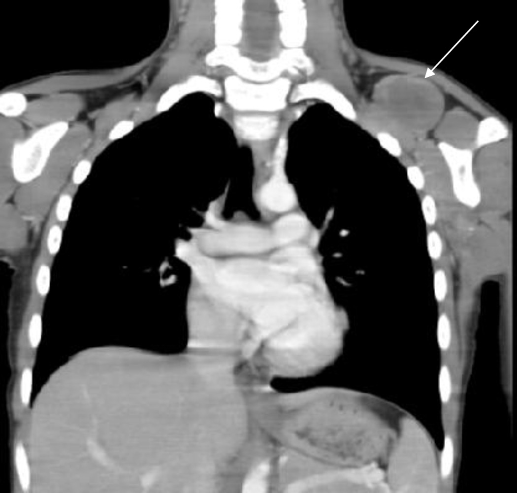
Results: Tumors were located in the extremities in nine patients, and one tumor each was located in the tongue and lung. Tumors were deep in seven patients and superficial in four patients. All tumors were well defined, solitary, and nonmetastatic at presentation. Tumors were complex solid-cystic in eight patients and completely solid in three patients. On T1-weighted images, all tumors had at least some areas hypointense to muscles, and six had a split-fat sign. On STIR or T2-weighted images, eight tumors had areas hypointense to adjacent muscle, and eight tumors had fluid signal intensity. On contrast-enhanced MRI studies, eight tumors had thick enhancing internal septations, and three had peripheral nodular gyriform enhancement. When we correlated imaging to pathologic findings, areas with hypointense signal intensity on both T1- and T2-weighted images were likely related to fibrous component; areas with fluid signal intensity on T2-weighted images were likely related to myxoid component. On CT, all four tumors were hypodense to muscle, and one tumor showed punctate calcific foci.
Conclusion: Low-grade fibromyxoid sarcoma is hypodense to muscle on CT. MRI may identify both fibrous and myxoid components of this rare pediatric soft-tissue sarcoma.
Keywords: ARST0332; CT; Children's Oncology Group; MRI; low-grade fibromyxoid sarcoma; pediatrics.
Figures


Similar articles
-
CT and MRI features of low-grade fibromyxoid sarcoma in the shoulder of a pediatric patient.Radiat Med. 2006 Aug;24(7):511-4. doi: 10.1007/s11604-006-0057-7. Radiat Med. 2006. PMID: 17058145
-
Imaging features of alveolar soft-part sarcoma: a report from Children's Oncology Group Study ARST0332.AJR Am J Roentgenol. 2014 Dec;203(6):1345-52. doi: 10.2214/AJR.14.12462. AJR Am J Roentgenol. 2014. PMID: 25415714 Free PMC article. Clinical Trial.
-
MRI of chondromyxoid fibroma.Acta Radiol. 2011 Oct 1;52(8):875-80. doi: 10.1258/ar.2011.110180. Epub 2011 Aug 11. Acta Radiol. 2011. PMID: 21835889
-
Magnetic resonance imaging of thymic epithelial tumors.Crit Rev Diagn Imaging. 1996 Aug;37(3):191-259. Crit Rev Diagn Imaging. 1996. PMID: 8872410 Review.
-
Peripheral primitive neuroectodermal tumor: dynamic CT, MRI and clinicopathological characteristics--analysis of 36 cases and review of the literature.Oncotarget. 2014 Dec 30;5(24):12968-77. doi: 10.18632/oncotarget.2649. Oncotarget. 2014. PMID: 25587032 Free PMC article. Review.
Cited by
-
Case report: Primary pleural low-grade fibromyxoid sarcoma in a 4-year-old boy with molecular confirmation.Front Oncol. 2023 Dec 20;13:1269078. doi: 10.3389/fonc.2023.1269078. eCollection 2023. Front Oncol. 2023. PMID: 38179169 Free PMC article.
-
Primary Osseous Low-grade Myxofibrosarcoma of Metatarsal Masquerading as Enchondroma: A Case Report.J Orthop Case Rep. 2022;12(5):35-39. doi: 10.13107/jocr.2022.v12.i05.2806. J Orthop Case Rep. 2022. PMID: 36660161 Free PMC article.
-
The role of molecular pathology in soft tissue tumor diagnosis: what the radiologist needs to know.Skeletal Radiol. 2025 Sep;54(9):1821-1840. doi: 10.1007/s00256-025-04934-1. Epub 2025 Apr 29. Skeletal Radiol. 2025. PMID: 40301146 Review.
-
Huge mesenteric low-grade fibromyxoid sarcoma: A case report and review of the literature.Rare Tumors. 2018 May 24;10:2036361318777031. doi: 10.1177/2036361318777031. eCollection 2018. Rare Tumors. 2018. PMID: 29854356 Free PMC article.
-
Case report: Primary pulmonary low grade fibromyxoid sarcoma progressing to dedifferentiation: probably due to TP53 driver mutation.Front Oncol. 2024 Mar 1;14:1329264. doi: 10.3389/fonc.2024.1329264. eCollection 2024. Front Oncol. 2024. PMID: 38496764 Free PMC article.
References
-
- Billings SD, Giblen G, Fanburg-Smith JC. Superficial low-grade fibromyxoid sarcoma (Evans tumor): a clinicopathologic analysis of 19 cases with a unique observation in the pediatric population. Am J Surg Pathol. 2005;29(2):204–210. - PubMed
-
- Evans HL. Low-grade fibromyxoid sarcoma: a clinicopathologic study of 33 cases with long-term follow-up. Am J Surg Pathol. 2011;35(10):1450–1462. - PubMed
-
- Folpe AL, Lane KL, Paull G, Weiss SW. Low-grade fibromyxoid sarcoma and hyalinizing spindle cell tumor with giant rosettes: a clinicopathologic study of 73 cases supporting their identity and assessing the impact of high-grade areas. Am J Surg Pathol. 2000;24(10):1353–1360. - PubMed
-
- Guillou L, Benhattar J, Gengler C, Gallagher G, Ranchere-Vince D, Collin F, et al. Translocation-positive low-grade fibromyxoid sarcoma: clinicopathologic and molecular analysis of a series expanding the morphologic spectrum and suggesting potential relationship to sclerosing epithelioid fibrosarcoma: a study from the French Sarcoma Group. Am J Surg Pathol. 2007;31(9):1387–1402. [Evaluation Studies Research Support, Non-U S Gov't]. - PubMed
-
- Doyle LA, Moller E, Dal Cin P, Fletcher CD, Mertens F, Hornick JL. MUC4 is a highly sensitive and specific marker for low-grade fibromyxoid sarcoma. Am J Surg Pathol. 2011;35(5):733–741. - PubMed
Publication types
MeSH terms
Substances
Grants and funding
LinkOut - more resources
Full Text Sources
Medical
