. 2016 Jul-Aug;37(4):207-15.
doi: 10.1097/BCR.0000000000000280.

Proteasome Inhibition After Burn Injury

Affiliations

Proteasome Inhibition After Burn Injury

P Geoff Vana et al. J Burn Care Res. 2016 Jul-Aug.

Abstract

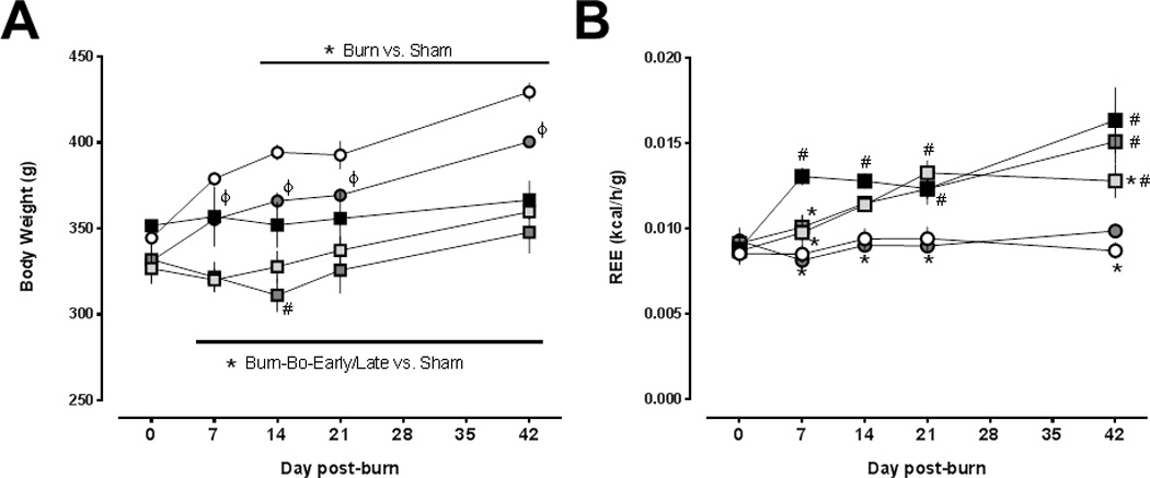
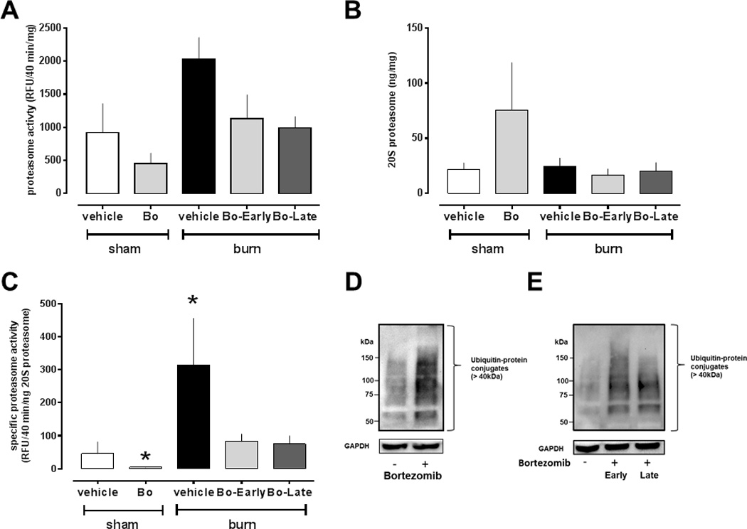
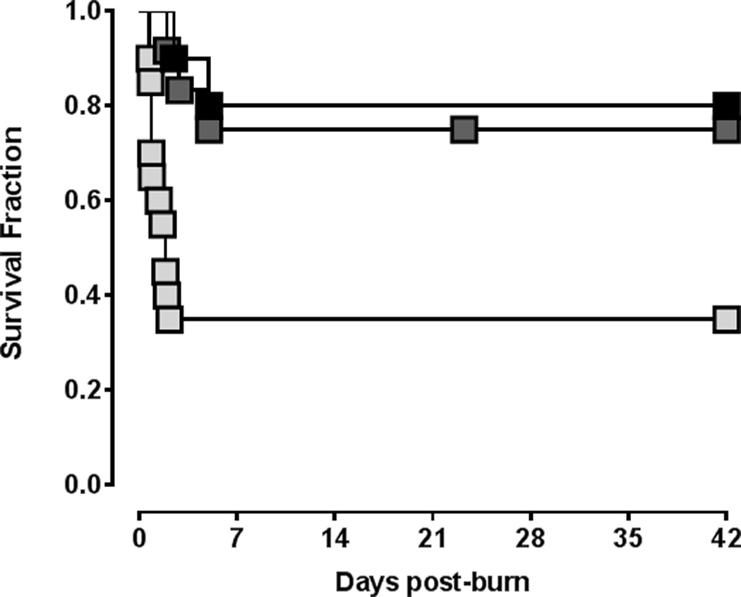
The objective of this study was to assess the effects of proteasome inhibition on the development of burn-induced hypermetabolism. Rats underwent 30-40% total BSA scald burn or sham injury. The proteasome inhibitor bortezomib (0.1 mg/kg) or vehicle (n = 10) was administered i.p. 3× weekly starting at 2 hours (early bortezomib, n = 20) or 48 hours (late-bortezomib, n = 13) postburn. Body weights were determined weekly. Resting energy expenditures (REE) were measured at days 0 (baseline), 7, 14, 21, and 42 postburn. At day 42, blood and pectoral muscle were harvested. Routine blood chemistry parameters were analyzed. Proteasome content, proteasome peptidase activities, and ubiquitin-protein conjugates were measured in muscle extracts. As compared with sham-vehicle-treated animals, specific proteasome activities were increased after burn and vehicle treatment. Bortezomib treatment inhibited proteasome activities and increased ubiquitin-protein conjugates after sham and burn injury. Bortezomib treatment did not affect REE after sham procedure. REE significantly increased by 47% within 7 days and remained elevated until day 42 after burn and vehicle treatment. After early-bortezomib treatment, burn-induced increases in REE were delayed and significantly reduced by 42% at day 42, as compared with vehicle treatment. With late-bortezomib treatment, burn-induced increases in REE were also delayed but not attenuated at day 42. Mortality was 20% with vehicle, 65% (median survival time: 1.875 days) with early-bortezomib and 25% with late-bortezomib treatment after burns (P < .05 early-bortezomib vs vehicle and late-bortezomib). Proteasome inhibition delays development of burn-induced hypermetabolism. Although proteasome inhibition early after burn injury reduces the hypermetabolic response, it significantly increases early burn-associated mortality.


Figures

Figure 1
Figure 1
(A) Body weights (g) after sham procedure and burn injury. Open circles: vehicle treatment – sham procedure (n=10). Grey circles: bortezomib treatment – sham procedure (n=12–13). Black squares: burn, vehicle treatment (n=8–10). Light grey squares: burn, early bortezomib treatment (n=7–20). Dark grey squares: burn, late bortezomib treatment (n=9–13). Data are mean ± SEM. ϕ p<0.05 sham procedure and vehicle treatment vs. sham procedure and bortezomib treatment. *: p<0.05 burn vs. sham procedure and vehicle treatment. #: p<0.05 burn and vehicle treatment vs. burn and late bortezomib treatment. (B) Resting energy expenditures (REE, kcal/h/g) after sham procedure and burn injury. Same symbols and sample sizes as in (A). Data are mean ± SEM. *: p<0.05 vs. burn and vehicle treatment. #: p<0.05 vs. sham procedure and vehicle treatment.
Figure 2
Figure 2
Proteasome peptidase activities, proteasome content and ubiquitin-protein conjugates in skeletal muscle extracts after sham procedure and burn injury (day 42). Sham procedure and vehicle treatment, n = 10. Sham procedure and bortezomib (Bo) treatment, n = 7. Burn and vehicle treatment, n=6. Burn, early Bo treatment, n = 6. Burn, late Bo treatment, n=6. (A) Proteasome peptidase activity (RFU/40 min/mg). (B) 20S proteasome concentration (ng 20S proteasome/mg protein). (C) Specific proteasome activity (RFU/40min/ng 20S proteasome). *: p<0.05 vs. sham procedure and vehicle treatment. (D) Top: Western blot analysis of ubiquitin-protein conjugates (>40kDa) in skeletal muscle extracts from uninjured animals after vehicle (−) and bortezomib (+) treatment. Bottom: Blots were re-probed with anti-GAPDH as a protein loading control. Migration positions of molecular mass standards are indicated on the left. (E) Top: Western blot analysis of ubiquitin-protein conjugates (>40kDa) in skeletal muscle extracts from animals after burn and vehicle (−) or early and late bortezomib (+) treatment. Bottom: Blots were re-probed with anti-GAPDH as a protein loading control. Migration positions of molecular mass standards are indicated on the left.
Figure 3
Figure 3
Survival after burn injury. Black squares: vehicle treatment (n=10). Light grey squares: Early bortezomib treatment (n=20). Dark grey squares: Late bortezomib treatment (n=13).

References

    1. Pereira C, Murphy K, Jeschke M, Herndon DN. Post burn muscle wasting and the effects of treatments. Int J Biochem Cell Biol. 2005;37(10):1948–1961. - PubMed
    1. Hart DW, et al. Determinants of skeletal muscle catabolism after severe burn. Ann Surg. 2000;232(4):455–465. - PMC - PubMed
    1. Garcia de Lorenzo y Mateos A, Ortiz Leyba C, Sanchez SM. Guidelines for specialized nutritional and metabolic support in the critically-ill patient: update. Consensus SEMICYUC-SENPE: critically-ill burnt patient. NutrHosp. 2011;26(Suppl 2):59–62. - PubMed
    1. Williams FN, Herndon DN, Jeschke MG. The hypermetabolic response to burn injury and interventions to modify this response. Clin Plast Surg. 2009;36(4):583–596. - PMC - PubMed
    1. Porter C, Hurren NM, Herndon DN, Borsheim E. Whole body and skeletal muscle protein turnover in recovery from burns. Int J Burns Trauma. 2013;3(1):9–17. - PMC - PubMed