

Herpes Simplex Virus Vector-Mediated Gene Delivery of Poreless TRPV1 Channels Reduces Bladder Overactivity and Nociception in Rats
- PMID: 26204493
- PMCID: PMC4651024
- DOI: 10.1089/hum.2015.026
Herpes Simplex Virus Vector-Mediated Gene Delivery of Poreless TRPV1 Channels Reduces Bladder Overactivity and Nociception in Rats
Abstract
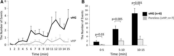
Increased afferent excitability has been proposed as an important pathophysiology of interstitial cystitis/bladder pain syndrome (IC/BPS) and overactive bladder (OAB). In this study, we investigated whether herpes simplex virus (HSV) vectors encoding poreless TRPV1, in which the segment in C terminus of TRPV1 receptor is deleted, suppress bladder overactivity and pain behavior using a rat model of chemical cystitis. Replication-defective HSV vectors encoding poreless TRPV1 were injected into the bladder wall of adult female Sprague-Dawley rats. Additionally, recombinant HSV virus (vHG) vectors were injected as control. Cystometry (CMG) under urethane anesthesia was performed 1 week after viral injection to evaluate bladder overactivity induced by resiniferatoxin (RTx, a TRPV1 agonist). RTx-induced nociceptive behavior such as licking (lower abdominal licking) and freezing (motionless head-turning) was observed 2 weeks after viral injection. GFP expression in L4/L6/S1 dorsal root ganglia and the bladder as well as c-Fos-positive cells in the L6 spinal cord dorsal horn were also evaluated 2 weeks after viral injection. In CMG, the poreless TRPV1 vector-treated group showed a significantly smaller reduction in intercontraction intervals and voided volume after RTx infusion than the vHG-treated control group. The number of the RTx-induced freezing events was significantly decreased in the poreless TRPV1 group than in the vHG group, whereas there was no significant difference of the number of RTx-induced licking events between groups. The number of c-Fos-positive cells in the DCM and SPN regions of the L6 spinal dorsal horn was significantly smaller in the poreless TRPV1 group than in the vHG group. Our results indicated that HSV vector-mediated gene delivery of poreless TRPV1 had a therapeutic effect on TRPV1-mediated bladder overactivity and pain behavior. Thus, the HSV vector-mediated gene therapy targeting TRPV1 receptors could be a novel modality for the treatment of OAB and/or hypersensitive bladder disorders such as IC/BPS.
Figures


Similar articles
-
Herpes simplex virus vector mediated gene therapy of tumor necrosis factor-α blockade for bladder overactivity and nociception in rats.J Urol. 2013 Jan;189(1):366-73. doi: 10.1016/j.juro.2012.08.192. Epub 2012 Nov 19. J Urol. 2013. PMID: 23174234
-
The effect of herpes simplex virus vector-mediated gene therapy of protein phosphatase 1α on bladder overactivity and nociception.Neurourol Urodyn. 2019 Feb;38(2):582-590. doi: 10.1002/nau.23882. Epub 2018 Nov 29. Neurourol Urodyn. 2019. PMID: 30499116
-
Effects of herpes simplex virus vectors encoding poreless TRPV1 or protein phosphatase 1α in a rat cystitis model induced by hydrogen peroxide.Gene Ther. 2018 Jan;25(1):20-26. doi: 10.1038/gt.2017.94. Epub 2017 Oct 20. Gene Ther. 2018. PMID: 29057994 Free PMC article.
-
Acute Intravesical Capsaicin for the Study of TRPV1 in the Lower Urinary Tract: Clinical Relevance and Potential for Innovation.Med Sci (Basel). 2022 Sep 10;10(3):50. doi: 10.3390/medsci10030050. Med Sci (Basel). 2022. PMID: 36135835 Free PMC article. Review.
-
Resiniferatoxin: Nature's Precision Medicine to Silence TRPV1-Positive Afferents.Int J Mol Sci. 2023 Oct 10;24(20):15042. doi: 10.3390/ijms242015042. Int J Mol Sci. 2023. PMID: 37894723 Free PMC article. Review.
Cited by
-
A bright future? Optogenetics in the periphery for pain research and therapy.Pain. 2018 Sep;159 Suppl 1(Suppl 1):S65-S73. doi: 10.1097/j.pain.0000000000001329. Pain. 2018. PMID: 30113949 Free PMC article. Review. No abstract available.
-
Nanotechnology for Pain Management: Current and Future Therapeutic Interventions.Nano Today. 2021 Aug;39:101223. doi: 10.1016/j.nantod.2021.101223. Epub 2021 Jun 19. Nano Today. 2021. PMID: 34899962 Free PMC article.
-
Next-generation replication-defective HSV vectors for delivery of large DNA payloads.Mol Ther. 2025 May 7;33(5):2205-2216. doi: 10.1016/j.ymthe.2025.03.055. Epub 2025 Apr 2. Mol Ther. 2025. PMID: 40181547 Review.
-
Gene Therapy Leaves a Vicious Cycle.Front Oncol. 2019 Apr 24;9:297. doi: 10.3389/fonc.2019.00297. eCollection 2019. Front Oncol. 2019. PMID: 31069169 Free PMC article. Review.
-
IPSE, a parasite-derived, host immunomodulatory infiltrin protein, alleviates resiniferatoxin-induced bladder pain.Mol Pain. 2020 Jan-Dec;16:1744806920970099. doi: 10.1177/1744806920970099. Mol Pain. 2020. PMID: 33342372 Free PMC article.
References
-
- Chancellor MB, Yoshimura N. Treatment of interstitial cystitis. Urology 2004;63:85–92 - PubMed
-
- Moutzouris D-A, Falagas ME. Interstitial cystitis: An unsolved enigma. Clin J Am Soc Nephrol 2009;4:1844–1857 - PubMed
-
- Lilly JD, Parsons CL. Bladder surface glycosaminoglycans is a human epithelial permeability barrier. Surg Gynecol Obstet 1990;171:493–496 - PubMed
-
- Slobodov G, Feloney M, Gran C, et al. . Abnormal expression of molecular markers for bladder impermeability and differentiation in the urothelium of patients with interstitial cystitis. J Urol 2004;171:1554–1558 - PubMed
-
- Ochs RL, Stein TW, Jr., Peebles CL, et al. . Autoantibodies in interstitial cystitis. J Urol 1994;151:587–592 - PubMed
Publication types
MeSH terms
Substances
Grants and funding
LinkOut - more resources
Full Text Sources
Other Literature Sources
Medical
