

S100B Up-Regulates Macrophage Production of IL1β and CCL22 and Influences Severity of Retinal Inflammation
- PMID: 26204512
- PMCID: PMC4512682
- DOI: 10.1371/journal.pone.0132688
S100B Up-Regulates Macrophage Production of IL1β and CCL22 and Influences Severity of Retinal Inflammation
Abstract
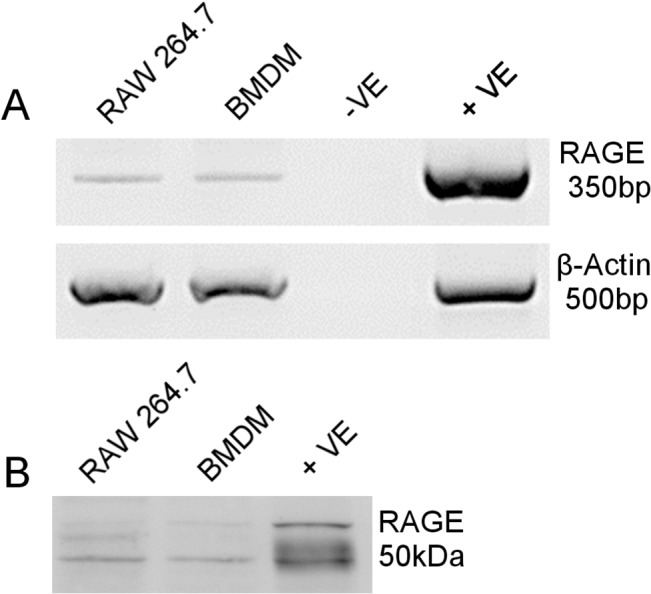
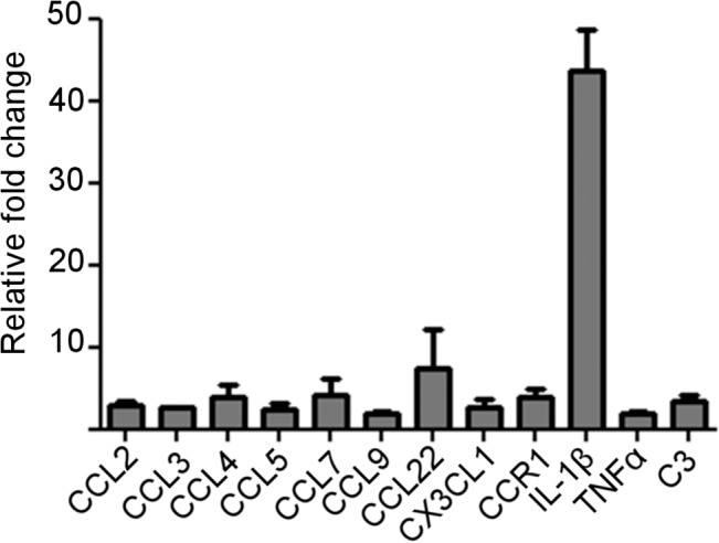
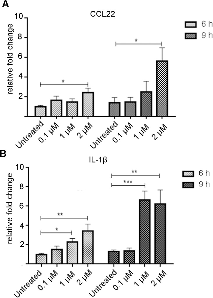
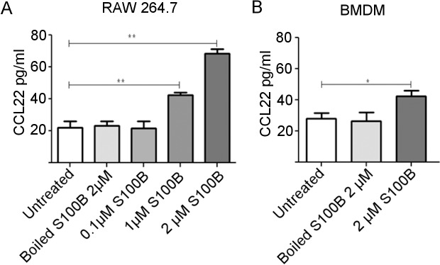
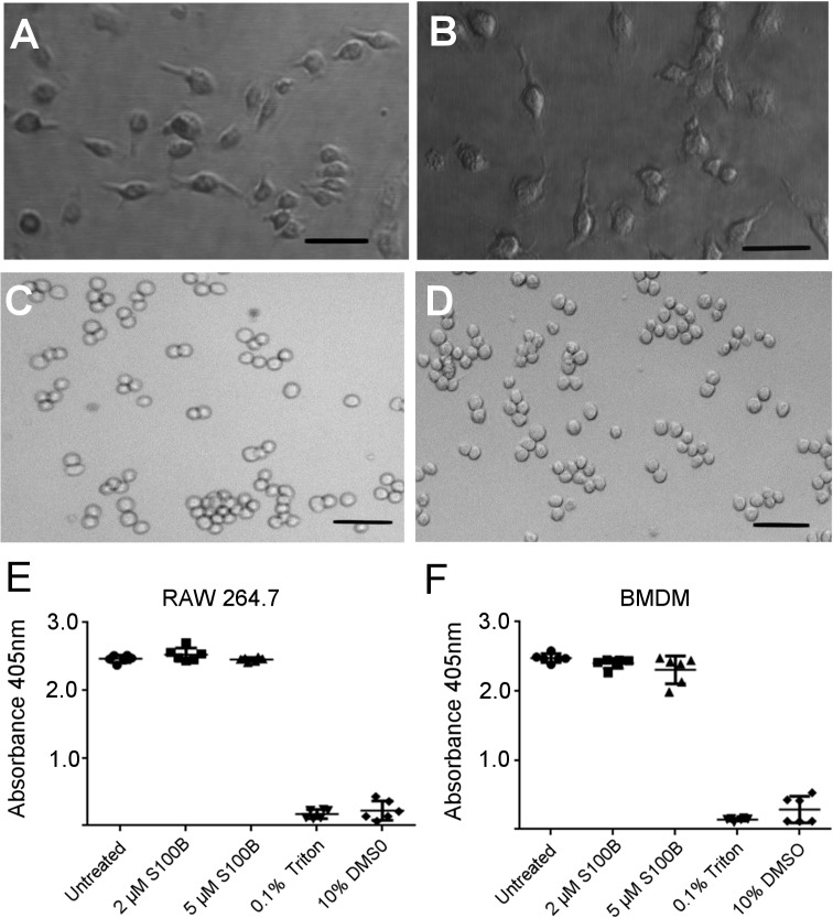
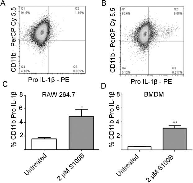
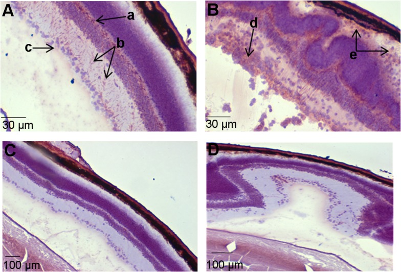
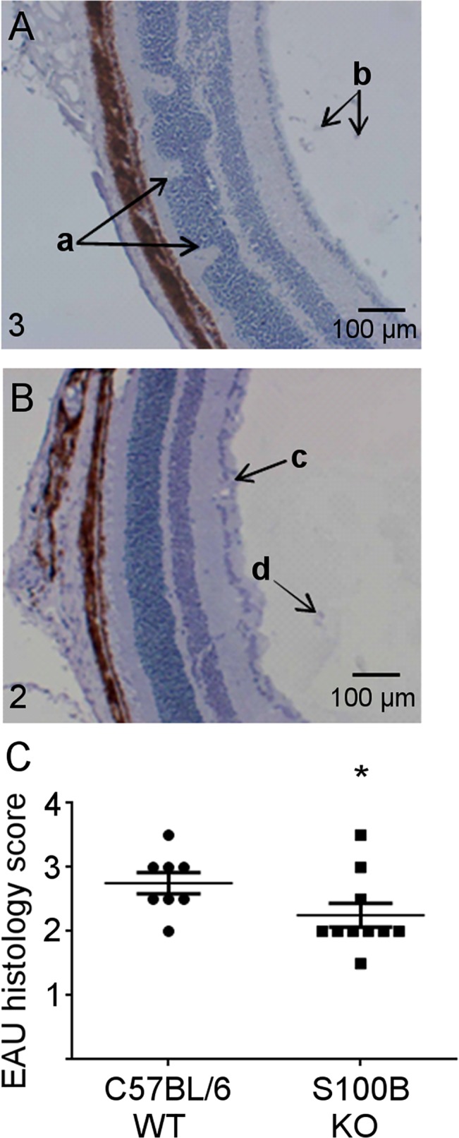
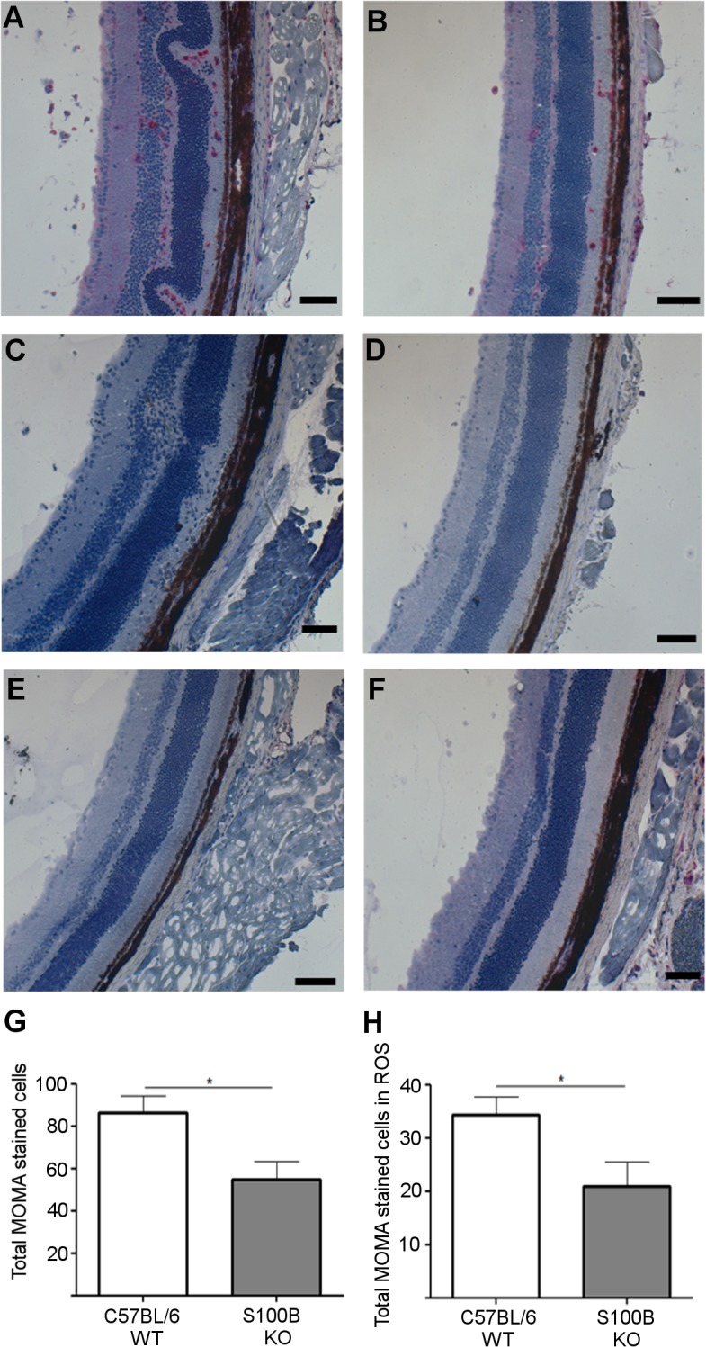
S100B is a Ca2+ binding protein and is typically associated with brain and CNS disorders. However, the role of S100B in an inflammatory situation is not clear. The aim of the study was to determine whether S100B is likely to influence inflammation through its effect on macrophages. A murine macrophage cell line (RAW 264.7) and primary bone marrow derived macrophages were used for in vitro studies and a model of retinal inflammatory disease in which pathogenesis is highly dependent on macrophage infiltration, Experimental Autoimmune Uveoretinitis, for in vitro study. Experimental Autoimmune Uveoretinitis is a model for the human disease posterior endogenous uveoretinitis, a potentially blinding condition, with an autoimmune aetiology, that mainly affects the working age group. To date the involvement of S100B in autoimmune uveoretinitis has not been investigated. Real-time PCR array analysis on RAW 246.7 cells indicated up-regulation of gene expression for various cytokines/chemokines in response to S100B, IL-1β and CCL22 in particular and this was confirmed by real-time PCR. In addition flow cytometry and ELISA confirmed up-regulation of protein production in response to S100B for pro-IL-1β and CCL22 respectively. This was the case for both RAW 264.7 cells and bone marrow derived macrophages. Induction of EAU with retinal antigen in mice in which S100B had been deleted resulted in a significantly reduced level of disease compared to wild-type mice, as determined by topical endoscopic fundus imaging and histology grading. Macrophage infiltration was also significantly reduced in S100B deleted mice. Real-time PCR analysis indicated that this was associated with reduction in CCL22 and IL-1β in retinas from S100B knock-out mice. In conclusion S100B augments the inflammatory response in uveoretinitis and this is likely to be, at least in part, via a direct effect on macrophages.
Conflict of interest statement
Figures

Similar articles
-
MicroRNAs in retina during development of experimental autoimmune uveoretinitis in rats.Br J Ophthalmol. 2016 Mar;100(3):425-31. doi: 10.1136/bjophthalmol-2015-306924. Epub 2015 Nov 5. Br J Ophthalmol. 2016. PMID: 26541434
-
Experimental autoimmune uveoretinitis (EAU)-related tissue damage and angiogenesis is reduced in CCL2⁻/⁻CX₃CR1gfp/gfp mice.Invest Ophthalmol Vis Sci. 2014 Oct 23;55(11):7572-82. doi: 10.1167/iovs.14-15495. Invest Ophthalmol Vis Sci. 2014. PMID: 25342612
-
The role of lipoprotein-associated phospholipase A2 in a murine model of experimental autoimmune uveoretinitis.PLoS One. 2015 Apr 15;10(4):e0122093. doi: 10.1371/journal.pone.0122093. eCollection 2015. PLoS One. 2015. PMID: 25874928 Free PMC article.
-
Caffeic acid phenethyl ester lessens disease symptoms in an experimental autoimmune uveoretinitis mouse model.Exp Eye Res. 2015 May;134:53-62. doi: 10.1016/j.exer.2015.03.014. Epub 2015 Mar 17. Exp Eye Res. 2015. PMID: 25795054
-
Macrophages and Uveitis in Experimental Animal Models.Mediators Inflamm. 2015;2015:671417. doi: 10.1155/2015/671417. Epub 2015 May 20. Mediators Inflamm. 2015. PMID: 26078494 Free PMC article. Review.
Cited by
-
S100b in acute ischemic stroke clots is a biomarker for post-thrombectomy intracranial hemorrhages.Front Neurol. 2023 Jan 23;13:1067215. doi: 10.3389/fneur.2022.1067215. eCollection 2022. Front Neurol. 2023. PMID: 36756347 Free PMC article.
-
Association of S100B polymorphisms and serum S100B with risk of ischemic stroke in a Chinese population.Sci Rep. 2018 Jan 17;8(1):971. doi: 10.1038/s41598-018-19156-w. Sci Rep. 2018. PMID: 29343763 Free PMC article.
-
Evolution of neuroinflammation across the lifespan of individuals with Down syndrome.Brain. 2020 Dec 1;143(12):3653-3671. doi: 10.1093/brain/awaa326. Brain. 2020. PMID: 33206953 Free PMC article.
-
Implication of Immunobiological Function of Melanocytes in Dermatology.Clin Rev Allergy Immunol. 2025 Mar 17;68(1):30. doi: 10.1007/s12016-025-09040-7. Clin Rev Allergy Immunol. 2025. PMID: 40097884 Review.
-
Negative regulators that mediate ocular immune privilege.J Leukoc Biol. 2018 Feb 12:10.1002/JLB.3MIR0817-337R. doi: 10.1002/JLB.3MIR0817-337R. Online ahead of print. J Leukoc Biol. 2018. PMID: 29431864 Free PMC article. Review.
References
-
- Zimmer DB, Cornwall EH, Landar A, Song W. The S100 Protein Family—History, Function, and Expression. Brain Research Bulletin 1995;37: 417–429. - PubMed
-
- Leclerc E, Fritz G, Vetter SW, Heizmann CW. Binding of S100 proteins to RAGE: An update. Biochimica et Biophysica Acta-Molecular Cell Research 2009;1793: 993–1007. - PubMed
-
- Donato R, Sorci G, Riuzzi F, Arcuri C, Bianchi R, Brozzi F, et al. S100B's double life: Intracellular regulator and extracellular signal. Biochimica et Biophysica Acta-Molecular Cell Research 2009;1793: 1008–1022. - PubMed
-
- Donato R. S100: a multigenic family of calcium-modulated proteins of the EF-hand type with intracellular and extracellular functional roles. International Journal of Biochemistry and Cell Biology 2001;33: 637–668. - PubMed
-
- Bierhaus A, Humpert PM, Morcos M, Wendt T, Chavakis T, Arnold B, et al. Understanding RAGE, the receptor for advanced glycation end products. Journal of Molecular Medicine-Jmm 2005;83: 876–886. - PubMed
Publication types
MeSH terms
Substances
LinkOut - more resources
Full Text Sources
Other Literature Sources
Molecular Biology Databases
Miscellaneous
