. 2015 Dec;22(12):1935-45.
doi: 10.1038/cdd.2015.99. Epub 2015 Jul 24.

miR-216a rescues dexamethasone suppression of osteogenesis, promotes osteoblast differentiation and enhances bone formation, by regulating c-Cbl-mediated PI3K/AKT pathway

Affiliations

miR-216a rescues dexamethasone suppression of osteogenesis, promotes osteoblast differentiation and enhances bone formation, by regulating c-Cbl-mediated PI3K/AKT pathway

H Li et al. Cell Death Differ. 2015 Dec.

Abstract

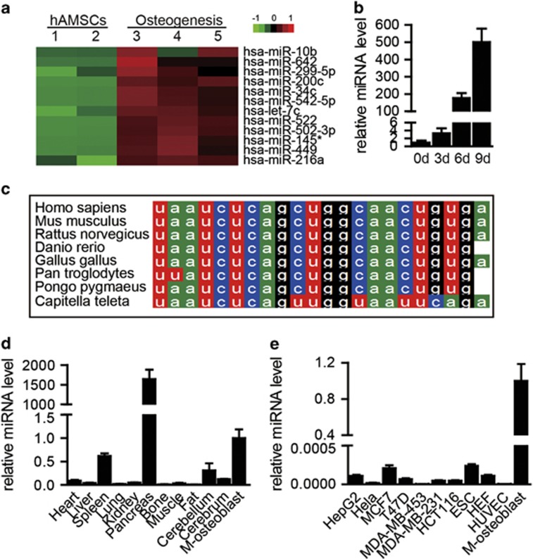
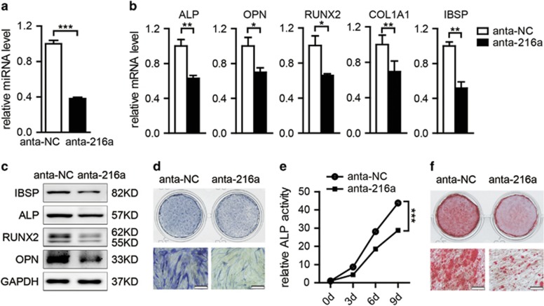
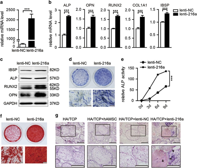
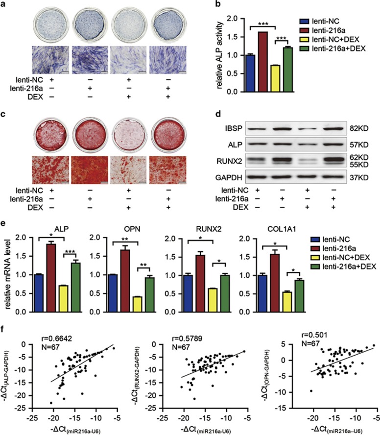
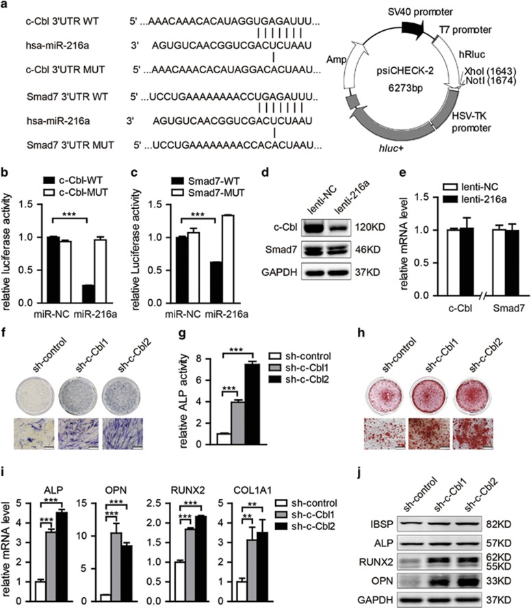
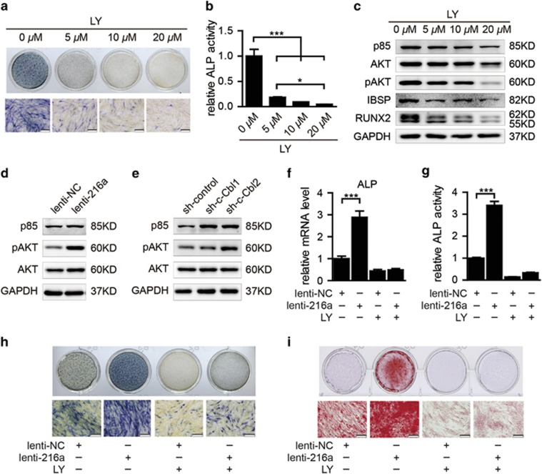
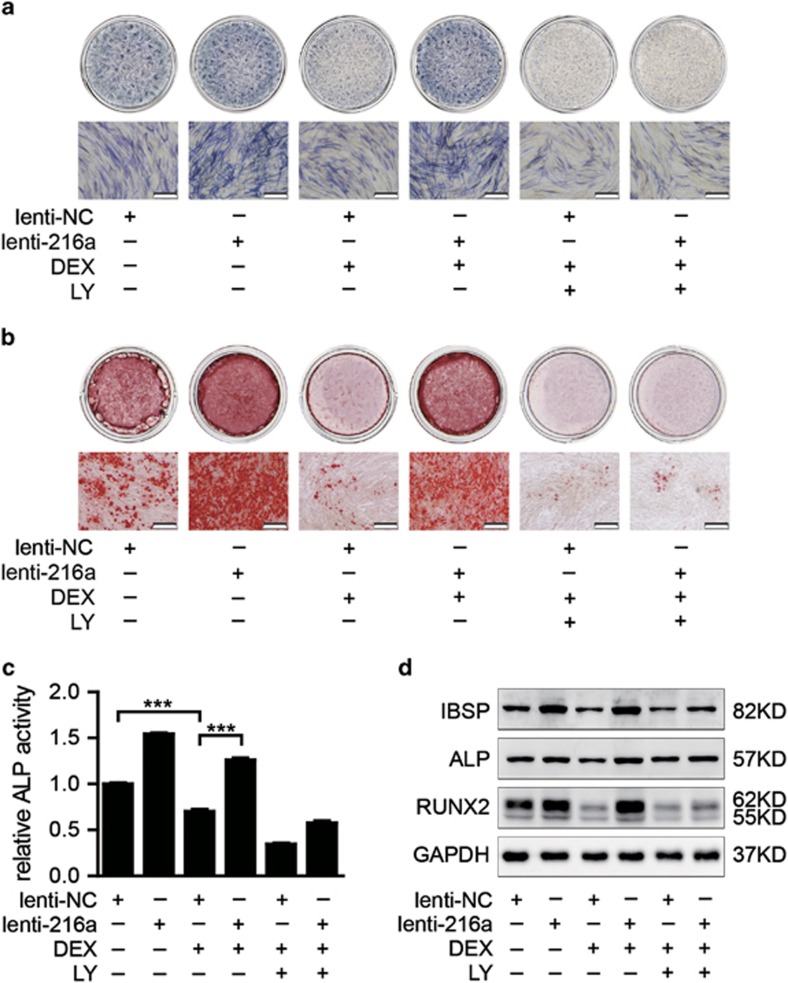
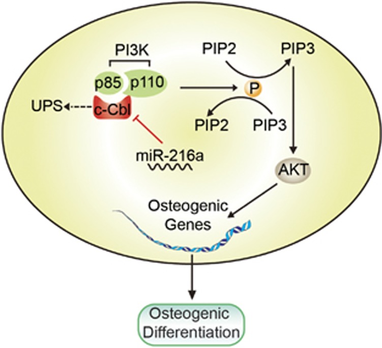
Osteoporosis is a disease marked by reduced bone mass, leading to an increased risk of fractures or broken bones. Bone formation is mediated by recruiting mesenchymal stem cells (MSCs). Elucidation of the molecular mechanisms that regulate MSC differentiation into osteoblasts is of great importance for the development of anabolic therapies for osteoporosis and other bone metabolism-related diseases. microRNAs (miRNAs) have been reported to have crucial roles in bone development, osteogenic differentiation and osteoporosis pathophysiology. However, to date, only a few miRNAs have been reported to enhance osteogenesis and regulate the suppressive effect of glucocorticoids on osteogenic differentiation. In this study, we discovered that miR-216a, a pancreatic-specific miRNA, was significantly upregulated during osteogenic differentiation in human adipose-derived MSCs (hAMSCs). The expression of miR-216a was positively correlated with the expression of bone formation marker genes in clinical osteoporosis samples. Functional analysis demonstrated that miR-216a can markedly promote osteogenic differentiation of hAMSCs, rescue the suppressive effect of dexamethasone (DEX) on osteogenic differentiation in vitro and enhance bone formation in vivo. c-Cbl, a gene that encodes a RING finger E3 ubiquitin ligase, was identified as a direct target of miR-216a. Downregulation of c-Cbl by short hairpin RNAs can mimic the promotion effects of miR-216a and significantly rescue the suppressive effects of DEX on osteogenesis. Pathway analysis indicated that miR-216a regulation of osteogenic differentiation occurs via the c-Cbl-mediated phosphatidylinositol 3 kinase (PI3K)/AKT pathway. The recovery effects of miR-216a on the inhibition of osteogenesis by DEX were attenuated after blocking the PI3K pathway. Thus, our findings suggest that miR-216a may serve as a novel therapeutic agent for the prevention and treatment of osteoporosis and other bone metabolism-related diseases.


Figures

Figure 1
Figure 1
Expression pattern of miR-216a. (a and b) The dynamic expression profile of miR-216a during osteogenesis in hAMSCs was detected by miRNA microarray and validated by TaqMan qRT-PCR. (c) Analysis of miR-216a conservation in mammals. (d and e) The tissue and cell line expression patterns of miR-216a compared with MSC-derived osteoblasts (M-osteoblast) were analyzed by TaqMan qRT-PCR. U6 was used as an internal control in the miRNA-specific TaqMan qRT-PCR. Quantitative data are presented as the mean±S.D. (n=3)
Figure 2
Figure 2
Downregulation of miR-216a inhibits the osteogenic differentiation of hAMSCs. (a) miR-216a expression was determined by TaqMan qRT-PCR in miR-216a antagomir (anta-216a)-transfected hAMSCs after 48 h compared with that in negative control antagomir (anta-NC)-transfected cells. (b and c) qRT-PCR and western blot analyses of the expression of osteoblast marker genes in anta-216a-transfected and anta-NC-transfected cells on day 6 of osteogenic differentiation. (d) ALP staining was performed on day 6 of osteogenic differentiation. (e) ALP activity was measured during osteogenic differentiation. (f) Alizarin red staining was performed to indicate mineral deposition on day 12. Scale bars: 200 μm. GAPDH was used as an internal control in the qRT-PCR and western blot analyses. U6 was used as an internal control in the miRNA-specific TaqMan qRT-PCR. Quantitative data are presented as the mean±S.D. (n=3). *P<0.05; **P<0.01; ***P<0.001 compared with the control
Figure 3
Figure 3
miR-216a promotes the osteogenic differentiation and bone formation of hAMSCs. (a) miR-216a expression was determined by TaqMan qRT-PCR in lenti-216a-infected hAMSCs compared with that in lenti-NC-infected cells. (b and c) qRT-PCR and western blot analyses of the expression of osteoblast marker genes in lenti-216a- and lenti-NC-infected cells on day 6 of osteogenic differentiation. (d) ALP staining was performed on day 6 of osteogenic differentiation. (e) ALP activity was measured during osteogenic differentiation. (f) Alizarin red staining was performed to indicate mineral deposition on day 12. (g) hAMSCs were infected with lenti-216a or lenti-NC and implanted into NOD/SCID mice. H&E staining was performed after 8 weeks of implantation. Arrowheads: bone matrix. Scale bars: 200 μm. GAPDH was used as an internal control in the qRT-PCR and western blot analyses. U6 was used as an internal control in the miRNA-specific TaqMan qRT-PCR. Quantitative data are presented as the mean±S.D. (n=3). ***P<0.001 compared with the control
Figure 4
Figure 4
miR-216a rescues the effect of DEX on osteogenic differentiation and is correlated with bone formation. (a and b) ALP staining and ALP activity detection were performed on day 6 of osteogenic differentiation. (c) Mineral deposition was indicated by Alizarin red staining on day 12. (d and e) Osteogenic differentiation was confirmed by qRT-PCR and western blot analyses of the osteogenic transcription factors and marker genes on day 6. (f) The correlation between the expression of bone formation makers and expression of miR-216a was analyzed in 67 osteoporosis samples. DEX: 10 nM DEX. Scale bars: 200 μm. GAPDH was used as an internal control in the qRT-PCR and western blot analyses. Quantitative data are presented as the mean±S.D. (n=3). *P<0.05; **P<0.01; ***P<0.001 compared with the control
Figure 5
Figure 5
c-Cbl is a direct target of miR-216a. (a) Computational analysis was performed for the complementarities of the miR-216a seed sequence to the 3'UTR of c-Cbl and Smad7. The WT or mutant-type (MUT) construct was inserted into the psiCHECK-2 reporter vector. (b and c) Luciferase activities were measured in the lysates, and the values were normalized to the psiCHECK vector and presented as the fold change of miR-NC. miR-216a: miR-216a mimics. (d) Western blot analysis of c-Cbl and Smad7 in hAMSCs after miR-216a overexpression. (e) qRT-PCR analyzed the expression of c-Cbl and Smad7 in hAMSCs after lenti-216a infection. (f and g) ALP staining and ALP activity detection were performed on day 6 of osteogenic differentiation in sh-c-Cbl1, sh-c-Cbl2 or sh-control-infected hAMSCs. (h) Alizarin red staining was performed to indicate mineral deposition on day 12. (i and j) The qRT-PCR and western blot analyses of the osteoblast transcription factors and marker genes were performed on 6 day of osteogenic differentiation. Scale bars: 200 μm. GAPDH was used as an internal control in the qRT-PCR and western blot analyses. All quantitative data are presented as the mean±S.D. (n=3). **P<0.01; ***P<0.001 compared with the control
Figure 6
Figure 6
PI3K pathway inhibition suppresses osteogenic differentiation and attenuates the enhancement of osteogenic differentiation by miR-216a overexpression. (a and b) PI3K inhibitor LY294002 was used to block the PI3K pathway. ALP staining and ALP activity detection were performed on day 6 of osteogenic differentiation in hAMSCs. (c) Western blot was performed to analyze the PI3K/AKT signaling pathway-related molecular elements and osteogenic factors after treatment with LY294002 on day 6. (d and e) Western blot was used to detect the protein levels of p85 and phospho-Akt after the overexpression of miR-216a or downregulation of c-Cbl in hAMSCs, respectively. (f) The ALP mRNA level was analyzed by qRT-PCR in induced cells on day 6. (g and h) ALP activity and ALP staining were performed on day 6. (i) Alizarin red staining was performed to indicate mineral deposition in induced cells after different treatments on day 12. LY: PI3K inhibitor LY294002. Scale bars: 200 μm. GAPDH was used as an internal control in the qRT-PCR and western blot analyses. All quantitative data are presented as the mean±S.D. (n=3). *P<0.05; ***P<0.001 compared with the control
Figure 7
Figure 7
PI3K pathway inhibition attenuates the recovery effects of miR-216a on the inhibition of osteogenesis by DEX. (a) ALP staining was performed on day 6 after different treatments. (b) Alizarin red staining was performed to indicate mineral deposition in osteogenesis induction of hAMSCs on day 12 after different treatments. (c) The ALP activity of cells was measured on day 6 of osteogenic differentiation. (d) Western blot was performed to analyze the protein level of osteogenic transcription factors and marker genes after different treatments on day 6 of osteoblast differentiation. LY: PI3K inhibitor LY294002. Scale bars: 200 μm. GAPDH was used as an internal control in the western blot analysis. All of the quantitative data are presented as the mean±S.D. (n=3). ***P<0.001 compared with the control
Figure 8
Figure 8
A schematic model for the miR-216a-mediated promotion of the osteogenic differentiation of hMSCs. miR-216a suppresses c-Cbl translation at the post-transcriptional level, resulting in the enhancement of the PI3K/AKT pathway and thereby promoting osteogenic differentiation

References

    1. Ahmed SF, Elmantaser M. Secondary osteoporosis. Endocr Dev 2009; 16: 170–190. - PubMed
    1. Canalis E, Delany AM. Mechanisms of glucocorticoid action in bone. Ann N Y Acad Sci 2002; 966: 73–81. - PubMed
    1. Yun SI, Yoon HY, Jeong SY, Chung YS. Glucocorticoid induces apoptosis of osteoblast cells through the activation of glycogen synthase kinase 3beta. J Bone Miner Metab 2009; 27: 140–148. - PubMed
    1. Pittenger MF, Mackay AM, Beck SC, Jaiswal RK, Douglas R, Mosca JD et al. Multilineage potential of adult human mesenchymal stem cells. Science 1999; 284: 143–147. - PubMed
    1. Zuk PA, Zhu M, Ashjian P, De Ugarte DA, Huang JI, Mizuno H et al. Human adipose tissue is a source of multipotent stem cells. Mol Biol Cell 2002; 13: 4279–4295. - PMC - PubMed

Publication types

MeSH terms