

doi: 10.1038/cr.2015.88.
Epub 2015 Jul 24.
Opposing roles of conventional and novel PKC isoforms in Hippo-YAP pathway regulation
Affiliations
- PMID: 26206313
- PMCID: PMC4528059
- DOI: 10.1038/cr.2015.88
Item in Clipboard
Opposing roles of conventional and novel PKC isoforms in Hippo-YAP pathway regulation
Cell Res.
2015 Aug.
No abstract available
Figures
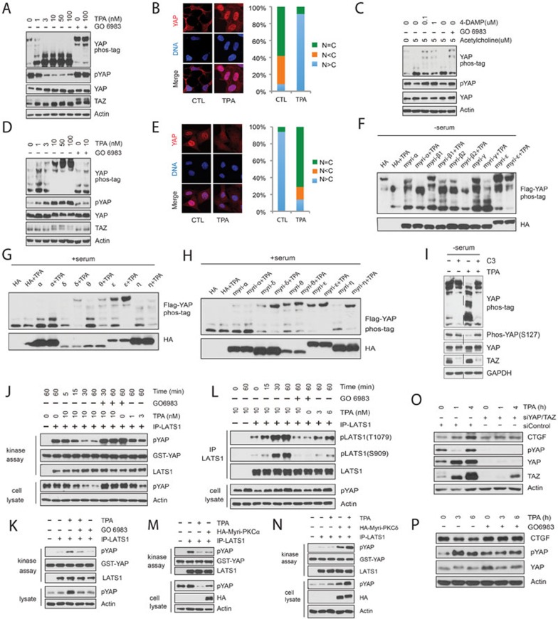
Opposing roles of conventional and novel PKC isoforms in Hippo-YAP pathway regulation. (A) TPA induces YAP and TAZ dephosphorylation in HEK293A cells. Cells were starved with serum-free medium for 12 h and treated with various amounts of TPA for 1 h. Where indicated, cells were pretreated with 200 nM PKC inhibitor GO6983 for 30 min. Cell lysates were analyzed by western blotting with indicated antibodies. Yap phosphorylation status was also determined by differential electrophoretic mobility shift on phos-tag-containing gels. (B) TPA promotes YAP nuclear accumulation in HEK293A cells. (C) Acetylcholine acts through muscarinic acetylcholine receptor M3 and PKC to activate YAP. Experiments were performed similarly as described in A. (D) TPA promotes YAP/TAZ phosphorylation in Swiss3T3 cells. Cells were cultured in serum-containing medium and stimulated with various doses of TPA for 1 h as indicated. (E) TPA promotes YAP cytoplasmic localization in MEF cells. (F) The conventional PKC promotes YAP dephosphorylation. 500 ng of individual HA-tagged myri-PKC isoforms and 2 ng of Flag-YAP were co-transfected into HEK293A cells. 24 h after transfection, cells were serum starved for 12 h and stimulated with 10 nM TPA for 1 h. (G, H) Expression of novel PKC enhances YAP phosphorylation in HEK293A cells. Indicated plasmids were co-transfected into HEK293A cells. 36 h later, cells were stimulated with fresh 10% serum-containing medium for 40 min followed by 10 nM TPA for 1 h. (I) Exoenzyme C3 prevents TPA-induced YAP dephosphorylation in HEK293A cells. 24 h after serum starvation, cells were pretreated with 2 μg/ml C3 for 4 h and then stimulated with TPA for 1 h. (J) TPA inactivates LATS in HEK293A cells. Serum-starved cells were stimulated with TPA as indicated. Endogenous LATS1 was immunoprecipitated for the kinase assay using GST-YAP as the substrate in vitro. Phosphorylation states of endogenous YAP in cell lysates were shown for comparison. (K, L) TPA activates LATS in Swiss3T3 cells. Experiments were performed similarly as described in J except that Swiss3T3 cells were not serum starved and that endogenous LATS1 was immunoprecipitated for immunoblotting with phosphor-LATS antibodies as indicated in L. (M) Constitutively active PKCα decreases LATS kinase activity. HA-Myri-PKCα was transfected into HEK293A cells as indicated. Cells were serum starved for 12 h and treated with 10 nM TPA for 1 h. Endogenous LATS1 was immunoprecipitated for kinase activity assay. (N) Constitutively active PKCδ promotes LATS activation. Plasmid encoding HA-Myri-PKCδ was transfected into HEK293A cells. 36 h after transfection, cells were first stimulated with 10% serum for 40 min and then with 10 nM TPA for 1 h. Endogenous LATS1 was immunoprecipitated for kinase assay. (O) TPA induces a YAP/TAZ-dependent CTGF accumulation in HEK293A cells. Control or YAP/TAZ-knockdown HEK293A cells were serum-starved for 12 h and then stimulated with TPA for various time points as indicated. Cell lysates were analyzed by immunoblotting. Notably, YAP phosphorylation recovered 4 h after TPA stimulation. This could be due to a transient LATS inhibition by TPA or a negative feedback induced by YAP/TAZ activation. (P) TPA decreases CTGF expression level in Swiss3T3 cells. Swiss3T3 cells were cultured in the presence of 10% serum for 12 h and then stimulated with TPA for the indicated time. Cell lysates were analyzed by immunoblotting.
References
-
- Wu S, Huang J, Dong J, Pan D. Cell. 2003. pp. 445–456. - PubMed
Publication types
MeSH terms
Substances
Grants and funding
LinkOut - more resources
Full Text Sources
Other Literature Sources
