. 2015 Jul 24;349(6246):428-32.
doi: 10.1126/science.aab1370.

INTRACELLULAR TRANSPORT. PI4P/phosphatidylserine countertransport at ORP5- and ORP8-mediated ER-plasma membrane contacts

Affiliations

INTRACELLULAR TRANSPORT. PI4P/phosphatidylserine countertransport at ORP5- and ORP8-mediated ER-plasma membrane contacts

Jeeyun Chung et al. Science. .

Abstract

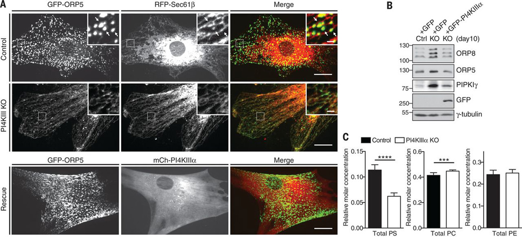
Lipid transfer between cell membrane bilayers at contacts between the endoplasmic reticulum (ER) and other membranes help to maintain membrane lipid homeostasis. We found that two similar ER integral membrane proteins, oxysterol-binding protein (OSBP)-related protein 5 (ORP5) and ORP8, tethered the ER to the plasma membrane (PM) via the interaction of their pleckstrin homology domains with phosphatidylinositol 4-phosphate (PI4P) in this membrane. Their OSBP-related domains (ORDs) harbored either PI4P or phosphatidylserine (PS) and exchanged these lipids between bilayers. Gain- and loss-of-function experiments showed that ORP5 and ORP8 could mediate PI4P/PS countertransport between the ER and the PM, thus delivering PI4P to the ER-localized PI4P phosphatase Sac1 for degradation and PS from the ER to the PM. This exchange helps to control plasma membrane PI4P levels and selectively enrich PS in the PM.


Figures

Fig. 1
Fig. 1. PI4P-dependent accumulation of ORP5 and ORP8 at ER-PM contact sites
(A) Confocal images of the middle or of the basal PM focal planes (illustrations at top) of HeLa cells expressing GFP-ORP5, GFP-ORP8L, or GFPORP8S either alone (left) or together with the PI4KIIIα complex (right). Scale bars, 10 µm. (B) Ratio of GFP fluorescence visible in the total internal reflection fluorescence (TIRF) (basal PM-associated fluorescence) versus epifluorescence fields (total fluorescence) of cells transfected as in (A) (mean ± SEM; P < 0.0001, t test, n = 8 to 20 cells). (C to E) Increase of ER-PM contacts produced by GFP-ORP5 expression as revealed with electron microscopy and accompanying morphometric analysis: contact length per unit PM length in (D) (mean ± SEM; P < 0.01, t test, n = 10 cells) and number of contacts per PM length in (E) (mean ± SEM; P < 0.001, t test, n = 10 cells). Scale bar, 200 nm. (F) Confocal live imaging of HeLa cells transfected with GFP-ORP5 and mCh-ORP8, showing partial recruitment of ORP8 under these conditions. Scale bar, 10 µm. (G) Co-immunoprecipitation of 3XFLAG-ORP8 with GFP-ORP5 in HeLa cells. Asterisk points to an nonspecific band. (H) Dissociation from the PM of GFP-ORP5 but not of mRFP-PHPLCδ upon acute treatment of cells with the specific PI4KIIIα inhibitor A1 (100 nM) induces. (I) ORP5 and ORP8 deletion constructs used for the experiments shown in (J). (J) Confocal live imaging of HeLa cells expressing the deletion constructs depicted in (I) and with a C-terminal GFP tag revealing that the PI4P-dependent PM recruitment of ORP5 and ORP8 is mediated by their PH domains. The smallest constructs show a partial accumulation in nuclei, as reported previously for other PH domains. Scale bars, 10 µm.
Fig. 2
Fig. 2. PI4KIIIα KO cells shows defects in PS metabolism
(A) Confocal live imaging of control (top) or PI4KIIIα KO MEFs (middle) coexpressing GFP-ORP5 and RFP-Sec61β showing ER-PM contacts at base of the cell and their absence in knockout cells. mCh-PI4KIIIα expression in the knockout cells rescues the phenotype (bottom). Regions enclosed by rectangles are shown at higher magnification in the insets. Scale bars, main fields, 10 µm; insets, 2 µm. (B) Immunoblot of the proteins indicated in lysates from control MEFs, PI4KIII knockout MEFs, and knockout MEFs expressing GFP-PI4KIIIα. (C) Lipidomics analysis of PI4KIIIα knockout and control MEFs (mean ± SEM; ****P < 0.0001, ***P < 0.001, t test, n = 3 cells).
Fig. 3
Fig. 3. Detection of PS and PIP in the ORD of ORP8
(A) Coomassie blue–stained SDS–polyacrylamide gel electrophoresis gel of purified 3XFLAG-ORDORP8 used for the mass spectrometry studies. (B) Mass spectrometry analysis of 3XFLAG-ORDORP8 showing the occurrence of apo and holo forms (with PS and PIP bound) in native conditions (bottom) and, for comparison, of the apo protein alone in denatured conditions (top). (C and D) Relative abundance of PS (C) and PIP (D) species (different acyl chain lengths) recovered after extraction from 3XFLAG-ORDORP8. (E) Sucrose-loaded heavy PC/PI4P liposomes (90:10 mol/mol, 2 mM lipids, 400 nm diameter) and light PC/PS liposomes (90:10 mol/mol, 2 mM lipids, 100 nm diameter) were incubated with no protein (–) or with 5 µM either WTORPORP8 or H514A, H515A mutant ORPORP8 for 15 min at 25°C. After centrifugation, supernatant (left) and pellet (right) fractions were collected, and the percentages of PI4P and PS recovered in the two fractions were assessed by means of high-performance liquid chromatography–based lipid analysis (mean ± SEM; ****P < 0.0001, ***P < 0.001, t test, n = 3 cells). Pellet fractions were normalized by PS recovery of (–) sample.
Fig. 4
Fig. 4. ORP5 and ORP8 mediate PS/PI4P exchange at the ER-PM contact sites
(A to D) Confocal live imaging of HeLa cells expressing iRFP-P4M [(A) and (B)] or GFP-evt2-2XPH [(C) and (D)] either alone (–) or together with WT or mutant ORP5, as indicated. The main fields show the fluorescence of iRFP-P4M and GFP-evt2-2XPH, respectively. The insets of (A) shows at high magnification the GFP-ORP5 fluorescence in the regions indicated in the main fields. The ratio of iRFP (A) or GFP (C) fluorescence visible in the TIRF versus epifluorescence fields is shown in (B) and (D) (mean ± SEM; P < 0.0001, t test, n = 14 to 25 cells). Scale bars, 10 µm; insets, 2 µm. (E) Effect of the double knockdown of ORP5 and ORP8 on the subcellular localization of the N-PH domain of ORP8. Scale bars, 10 µm. (F) Confocal live microscopy showing that recruitment of ΔPH-ORP5 to the PM with 1 µm rapamycin induces dissociation of iRFP-P4M. Scale bar, 10 µm for the main fields; 2 µm for the time sequence. (G to L) Quantification of fluorescent signals in the TIRF fields upon rapamycin-induced PM recruitment of FKBP12 fused to the ΔPH-ORP proteins (mean ± SEM). (G) Loss of iRFP-P4M and increase of GFP-evt2-2XPH upon recruitment of ΔPH-ORP5WT (n = 6 cells). (H) Loss of iRFP-P4M, but not of GFP-PH-PLCδ, upon recruitment of ΔPH-ORP5WT (n = 8 cells) (I) Loss of iRFP-P4M upon ΔPH-ORP5WT or ΔPH-ORP8WT recruitment, but not upon recruitment of ΔPH-ORP5H478/ 479A or ΔPH-ORP5L389D (n = 6 to 10 cells). (J) GFP-evt2-2XPH fluorescence upon recruitment of ΔPH-ORP5WT ΔTM (blue, n = 18 cells) or ΔPH-ORP5WT pretreated with 100 nM A1 (10 min) (green, n = 10 cells) (K) Loss of iRFP-P4M only if the ΔPH-ORPWT construct is tethered to the ER by its transmembrane region (n = 4 cells). (L) The knockdown of Sac1 (inset, immunoblot) impairs the decrease of iRFP-P4M fluorescence upon recruitment of ΔPH-ORP5WT (n = 6 to 9 cells).

References

    1. Stefan CJ, Manford AG, Emr SD. Curr. Opin. Cell Biol. 2013;25:434–442. - PMC - PubMed
    1. Holthuis JCM, Menon AK. Nature. 2014;510:48–57. - PubMed
    1. Moser von Filseck J, Mesmin B, Bigay J, Antonny B, Drin G. Biochem. Soc. Trans. 2014;42:1465–1470. - PubMed
    1. Toulmay A, Prinz WA. Curr. Opin. Cell Biol. 2011;23:458–463. - PMC - PubMed
    1. Levine T, Loewen C. Curr. Opin. Cell Biol. 2006;18:371–378. - PubMed

Publication types

Substances

LinkOut - more resources